Abstract
The term “geriatric” refers to old age. Determining if a bird is geriatric is based on the species’ average life expectancy. Up until more recently, there have not been enough geriatric birds of most commonly kept species available, either wild-caught or raised in captivity, to be able to study them; therefore, it had not been known at what age changes in their physical and mental health begin to occur. Although tables with lifespans have been published, other factors influence lifespans, such as nutrition, genetics, and exercise. These can either accelerate or delay changes related to aging. Consequently, geriatric, as it relates to birds, is the age at which medical conditions associated with aging are being documented and reported. As they age, birds will suffer from many of the same ailments that humans and other mammals do. Avian veterinarians are seeing more and more geriatric birds in their practices, as the larger birds which were purchased in the 1980’s and 1990’s reach the end of their lifespans, and the smaller birds, living longer than they had in the past, reach the end of their lifespans also. The illnesses they develop affect their behavior and mental states.
Introduction
The pet trade of the 1970’s and 1980’s brought to the United States and some European countries huge numbers of wild-caught birds, and their progeny live on today. Companion birds now live in nearly every country of the world. In spite of this, there is very little information available on the geriatric parrot. So little was known about these birds in the 1970’s,1980’s, and 1990’s that veterinarians scrambled to learn as much as possible, as quickly as possible, to assist the owners and their birds. As time went on, organizations such as the Association of Avian Veterinarians (AAV) were formed for just that purpose, and veterinarians began to specialize in avian medicine. Still, even today there is a dearth of specialists. Many countries, and even some states in the U.S. have no avian veterinarians at all.
Now, forty to fifty years after the huge influx of exotic birds, clinicians are dealing with both wild-caught and captive-bred birds which are reaching old age. One of the complications of caring for both older imported and captive-bred birds is that their health issues may not be the same; in some cases, they must deal with conflicting data.
Avian veterinarians are seeing more and more geriatric birds these days, and the birds are falling victim to the same types of illnesses that aging humans do. These illnesses affect their behavior and mental states, just as they do in humans. Malnutrition, disease, and poor husbandry have caused untold numbers of imported birds to die in captivity instead of reaching their expected lifespans.
The average lifespan of a bird is the life expectancy for that particular species. Longevity refers to the maximum lifespan that can be expected under ideal conditions. Many birds never reach their maximum lifespans, nor even get close to reaching them. “The bird’s lifespan is affected by its species, genetic inbreeding, size, concurrent disease, diet, and environment.” 20 If the diseases they suffered from had been caught earlier, many of the birds would have had longer lifespans. 44
1 Senescence
Senescence refers to the aging process in an organism. Researchers have difficulty identifying the true onset of aging in psittacines because it is so subjective. Years have to pass during which several generations of captive-bred birds of each species have been followed in order to determine the rates of senescence in these species. 28
Since birds are living longer, the incidence of geriatric-onset diseases has increased. Information on the physical, emotional, mental, and behavioral issues related to senescence will guide both practitioner and owner in dealing with age-related illness. Clinicians must deal not only with obvious illnesses, but with other issues as well: 28
- They must work with the client to preserve functional ability and mobility.
- They need to recognize and provide supplemental-care needs.
- They need to communicate with the client any quality-of-life/end-of-life concerns.28
1.1 The Effect of Social Isolation on Senescence: A Study
When considering the effect that socialization has on the lifespans of birds, one study is worth discussing. This study was done by D. Aydinonat, et al. in 2014 to determine the effect of social isolation on senescence in parrots. “Telomeres, parts of some chromosomes, control chromosome stability and cellular aging, but aging and exposure to chronic stress are thought to cause a change of the telomere length. The study was conducted to determine the effect of social isolation on telomere length in the highly social and intelligent African Grey parrot (Psittacus erithacus erithacus). The study population of captive birds consisted of 26 single-housed birds and 19 pair-housed birds between 9 months and 45 years of age.” 1 PCR testing was used to measure the telomere length. They found that telomere length declined with age, and socially isolated parrots had significantly shorter telomeres compared to pair-housed birds, even among birds of similar ages. “This study supports the hypothesis that telomeres provide a biomarker indicating exposure to chronic stress.” 1 Many of our socially isolated birds experience stress on a daily basis, and the older the bird, the shorter the telomeres became, indicating that chronic stress shortens the lives of birds. 1


Images 1, 2 This 92-year-old parrot was passed down through several generations in the same family (images courtesy Hub Pages; http://hubpages.com/animals/From-the-parrots-beak-910-edition).
1.2 Rate of Senescence in Birds
Bird species are strikingly long-lived compared to humans and other animals. “Many species live up to three times longer than mammals of equal body mass. The slow rates of senescence experienced by birds is a paradox.
- They have very high metabolic rates, high body temperatures, and blood-glucose levels.
- Their blood glucose levels are two to four-times greater than mammals.
- Their lifetime energy expenditures are up to 15 times higher than those of mammals.
According to current biochemical theories of aging, birds should experience accelerated tissue damage as a result of the damaging byproducts of oxidation.” 2, 29 Instead, birds age more slowly and gradually because of adaptations they have developed for preventing tissue damage due to oxidation and “their unusual capacity for neuroregeneration in the brain.” 2,28,29 The oxidative-damage theory suggests that “Species who are oxygen-reactive during normal oxidative metabolism processes undergo molecular changes and age-related declines. The fact that birds age slowly, despite high energy expenditures over a lifetime, suggests that birds possess special adaptive defenses against oxidative damage.”29 These adaptations allow birds to age very slowly until the end time of their lives. At that point, senescence occurs quickly.
This slower rate of senescence in birds has been documented with data from those in charge of zoo populations, veterinarians who see pet birds, and aviculturists who have tagged and recaptured wild birds and followed them throughout their lives. Birds maintained in captivity experience age-related changes that are not all that different from those in mammals; these changes just occur later and more gradually in birds. 28
2 Lifespan and Longevity
The average lifespan of a bird is the life expectancy for that particular species. Longevity refers to the maximum lifespan that can be expected under ideal conditions. Many birds never reach their maximum lifespans nor even get close to reaching them. According to C. Greenacre, “The lifespan of a bird depends on its species, genetic inbreeding, size, concurrent disease, diet, and environment.”20 Some of the more commonly seen geriatric diseases and conditions are arthritis, neurological disease, heart disease, neoplasia, obesity, atherosclerosis, chronic malnutrition, chronic aflatoxin exposure (carcinogenic mycotoxins, or mold-spore diseases), and cataracts. If these diseases had been caught earlier, many of the birds would have had longer lifespans. 44
Until recently, geriatric medicine has been a neglected area of avian medicine. Infectious diseases, inadequate diets, and poor care meant that most pet birds did not live long enough to develop geriatric conditions. However, as the knowledge base of avian medicine has increased, so have the lifespans of pet birds. S. Austed states that, “Most pet birds have the potential to live 20-80 years, depending on their size (larger birds live longer than smaller ones), diet (herbivores live longer than carnivores or omnivores), and socialization (highly social species live longer than solitary species).” 3
2.1 The Effect of Flight on Lifespans
In birds, species that are capable of flight live considerably longer than non-flying species of similar body size. “This could be because flight enables animals to escape some of the environmental dangers they encounter, such as predators or destruction or deterioration of their habitats. In addition, birds that are weak flyers are thought to age more rapidly than stronger-flying species. Birds of the order Galliformes (pheasants, quail, chickens, turkeys) are weak fliers, and they appear to be exceptionally short-lived compared to strong fliers.” 2

Image 3. A 20-year-old lutino cockatiel is shown after having the jugular vein moistened with alcohol for venipuncture. At this age, even minor restraint has caused the bird to appear listless and sleepy. Geriatric birds require more gentle handling and for shorter periods of time (image courtesy Doneley, Harrison, Lightfoot; used with permission). 16
FREE Training to Stop Your Bird's Biting
Get FREE access to Beak School – the only system designed to train your bird. Stop behavioral problems like biting, screaming, feather plucking.
Get the Free Training2.2 Geriatric Ages and Maximum Lifespans of Commonly Kept Companion Birds
This is a most interesting study. The following table comes from Clinical Avian Medicine, published in 2006. At that time, pet birds were not expected to live as long as their wild counterparts. Although the maximum lifespans are long, the actual average lifespan was much shorter. The Geriatric-in-Years data below was not only the period at which the birds were considered geriatric, it was also the average lifespan of each species. Very few birds reached their maximum lifespans until recently. You will also note that information is only provided for 14 species; in the case of some of the very long-lived species, there was no information available at the time, indicating that very few birds had reached geriatric status so that research data could have been obtained.
|
Species |
Geriatric Ages/Average Lifespans in Years |
Maximum Lifespans In Years |
|
Budgerigar (Melopsitticus undulatus) |
6-12 |
18 |
|
Cockatiels (Nymphicus hollandicus) |
12-18+ |
32 |
|
Sun conure (Aratinga solstitialis) |
18-25 |
25 |
|
Green-cheeked conure (Pyrrhura molinae molinae) |
12-15 |
25 |
|
Peach-faced lovebird (Agapornis roseicollis) |
10-15 |
12 |
|
Yellow-naped Amazon (Amazona ochrocephala auropalliata) |
35-45 |
* |
|
Blue-fronted Amazon (Amazona aestiva) |
25-35 |
80 |
|
Congo grey (Psittacus erithacus) |
20-25 |
50 |
|
Eclectus parrot (Eclectus roratus) |
15-20 |
20 |
|
Galah (rose-breasted cockatoo) (Eolophus roseicapillus) |
18-20 |
20 |
|
Umbrella cockatoo (Cacatua alba) |
20? |
* |
|
Moluccan cockatoo (Cacatua moluccensis |
25? |
* |
|
Yellow-collared macaw (Ara auricollis) |
22-27 |
* |
|
Blue and gold macaw (Ara ararauna) |
30-40 |
50 |
|
Green-winged macaw (Ara chloroptera) |
35-45 |
* |
*No data available (chart courtesy L. Wilson, P. Linden, and T. Lightfoot).61
Compare the data in the above chart with the one below. You will note that the species above for which no data was available have data listed in the table below. And the species with data in the above table show a much longer life-expectancy in the table below. This is attributed to:
- The improved education of veterinarians, both in veterinary school and on-going education of those in practice
- Education of the clients by more knowledgeable veterinarians
- Introduction of pellets as part of a balanced diet (At that time, pellets were considered the best way to improve the health of birds captured from the wild and cage birds in general. Today, that thinking has been replaced by the recommendation to feed a well-rounded, nutritious diet without pellets. See Reference Number 61 for a paper on that subject.)
- Improved husbandry, and
- Awareness of the physical, social, mental and emotional needs of these birds.
These are not the maximum life expectancies, but the average life expectancies. Maximum life expectancies would be far greater. For example, the cockatiel’s average life expectancy is only 10-15 years, but the maximum, being held now by many birds, is 32 years of age, and one bird has been anecdotally reported to be 34 years of age.
To add another perspective, Petrak and Minsky, in their 1982 text, note that they had rarely seen budgies over 10 or 11 years of age or canaries over 12 or 14 years of age;52 however, in the above chart, the budgie’s maximum life span is 18 years.
2.3 Life Expectancy of Some Common Companion Bird Species as of 1982 15
Species Average Life Expectancy in Years
|
African Grey, Congo |
50-60 |
|
African Grey, Timneh |
25-35 |
|
Alexandrine parrot |
25-35 |
|
Amazon, blue-crowned |
50-70 |
|
Amazon, blue-fronted |
50-70 |
|
Amazon, double yellow-headed |
50-70 |
|
Amazon, yellow-naped |
50-70 |
|
Budgerigar/parakeet |
7-12 |
|
Caique |
30-40 |
|
Canary |
6-12 |
|
Cockatiel |
10-15 |
|
Cockatoo, bare-eyed (little corella) |
30-40 |
|
Cockatoo, greater sulphur crested |
50-70 |
|
Cockatoo, lesser sulphur-crested |
40-60 |
|
Cockatoo, Major Mitchell |
40-60 |
|
Cockatoo, medium sulphur-crested |
40-60 |
|
Cockatoo, rose-breasted (galah) |
20-40 |
|
Cockatoo, citron-crested |
50-60 |
|
Cockatoo, Moluccan |
50-60 |
|
Cockatoo, Triton |
50-60 |
|
Cockatoo, umbrella |
50-60 |
|
Conure, Aratinga spp. |
15-25 |
|
Conure, Pyrrhura spp. |
15-25 |
|
Eclectus, red -sided |
25-30 |
|
Indian Ring-neck |
25-30 |
|
Jardine parrot (red-fronted |
20-25 |
|
Loriket, rainbow |
15-20 |
|
Lory, chattering |
30-35 |
|
Lovebird |
7-15 |
|
Macaw, blue and gold |
50-80 |
|
Macaw, green-winged |
60-90 |
|
Macaw, Hahn’s |
25-30 |
|
Macaw, hyacinth |
60-90 |
|
Macaw, military |
50-60 |
|
Macaw, scarlet |
50-80 |
|
Meyer’s parrot |
25-35 |
|
Mynah bird |
12 |
|
Pionus, blue-headed |
20-25 |
|
Pionus, white-capped |
25-30 |
|
Quaker (monk parakeet) |
20-30 |
|
Senegal parrot |
15-25 |
(Chart courtesy B. Doneley) 15
This data is, by its nature, subjective, depending on the methods used in research, the number of birds monitored, how many years they are monitored, and whether the birds were from zoos, aviaries, or private homes.
2.4 Lifespan study in 2014
In a 2014 study was complete by the group then known as the International Species Information System (ISIS), and now known as Species360. This organization receives its information from zoos around the world and examines parrots’ life history records. In this study, 83,212 parrots’ life history records were examined. The following lifespan information of several species was extrapolated: 20
Species Average lifespan Approximate Longevity Record
|
Budgie |
5-7 years |
18 years |
|
Cockatiel |
5-7 years |
32 years |
|
Lovebird |
10 years |
13-34 years |
|
Conure |
20 years |
6-60 years |
|
Amazon |
15-50 years |
22-66 years |
|
African Grey |
15-40 years |
48-60 years (92) |
|
Cockatoo |
15-30 years |
27-92 years |
|
Macaw |
15-30 years |
32-63 years |
|
Lory |
7 years |
17-30 years |
(Chart courtesy C. Greenacre) 20
It is most interesting that these average lifespans are generally the same in all charts; however, there is a huge discrepancy in the maximum lifespans due to the number of variables involved. (Approximate Longevity Record).
2.5. Extended Lifespan of Birds Today
Margaret Wissman, in her article, Growing Old Gracefully, discusses Icarus, a double-yellow-headed Amazon parrot which came to her for his yearly examination. “He was a large, handsome bird, with sleek, shiny feathers. He would flare his tail and ruffle up his head feathers, saying ‘hello’ and ‘here’ while pacing back and forth in his travel cage. This healthy bird represents a new class of birds being seen more and more frequently in avian practices today. Icarus is 24 years old and considered a middle-aged bird.” 64
She states that advances in avian medicine and improved husbandry practices are causing avian patients to living longer. She routinely sees cockatiels in their twenties, mid-sized birds in their teens, and macaws in their forties and fifties. Amazons can be very long-lived. In her own collection, she had, at the time of this writing, a double yellow-headed Amazon patient, Rocky, that was over 36 years old. The owners have proof of his age; he was purchased with adult head coloration in 1962. Even though budgies tend to have tumors and for the most part live only until 10 years of age, she is seeing them now living into their teens. Today, because of leg banding and more accurate breeder records than in the past, the ages of many domestically hatched pet birds can be tracked. 64
2.6 Clues for Determining the Age of a Bird
Although it may be difficult or impossible to pin down an older bird’s age, there are some clues that can be used to estimate the age of birds. Old birds may suffer from:
- Muscle wasting and weight loss.
- Limited range-of-motion; some birds may only be able to adequately move their wings and legs.
- Changes in skin appearance in birds with bare facial skin; changes include as wart-like blemishes, cysts and wrinkling.
- Depigmentation on the skin of the feet in spots.
- Diminished feather luster. 64
Some owners insist that they know the age of an imported bird, possibly due to information given at the time of purchase. If the birds were imported as babies, it is possible to determine the age, but for the most part, birds imported as adults cannot be given a numerical age since age changes cannot be predicted. 64
3 Immunosenescence – The Effect of Aging on the Immune System
An animal’s immune function declines with age, according to H. Pendle. As animals age, “Important cell populations are not fully replenished, and eventually this affects its immune function. Important T-cell and B-cell numbers decline as these organs decline, and this has the greatest impact on the immune system. Researchers hypothesize from this that the body reallocates its energy and nutrient supplies to the less costly immune responses so that the body may use its resources for overall biologic fitness in older birds.” 51
4 Diseases and Conditions Frequently Found in Geriatric Birds.
4.1 Percentage of Elderly Birds Suffering from Specific Diseases
In 2009, Zoo/Exotic Pathology Service (ZEPS) released a summary of disease conditions found in older psittacines. The population chosen included commonly kept species. In their report, budgerigars were considered elderly at 6 years; cockatiels at 12 years; and large psittacines, such as Amazons, macaws, cockatoos, and African greys, at 30 years. The table provides the name of each species and the disease conditions most frequently seen in that species. 54 The number of birds studied and the age ranges are:
African greys: 41 studied, 30-53 years
Amazons: 168 studied, 30-86 years
Budgies: 229 studied, 6-15 years
Cockatiels: 383 studied, 12-30 years
Cockatoos 27 studied, 30-45 years
Lovebirds: 206 studied, 6-18 years
Macaws: 66 studied, 30-60 years 54
4.2 Diseases of Old Age in Commonly Kept Species
|
Species |
Tumors |
Liver Disease |
Inflammatory Skin Lesions |
Heart Lesions |
Gonadal Degeneration |
|
Budgerigar |
66.8% |
5.7% |
3.5% |
3% |
4.4% |
|
Lovebird |
34.4% |
10% |
22.8% |
11.2% |
5.3% |
|
Amazon |
35% |
8% |
5.9% |
8.9% |
3% |
|
Cockatiel |
49.6% |
10% |
7.8% |
8.6% |
2.3% |
|
Macaw |
37.8% |
7.5% |
6% |
15% |
0 |
|
Cockatoo |
29.6% |
3.7% |
11% |
7.4% |
0 |
|
African grey |
34% |
7.3% |
2.4% |
19.5% |
0 |
|
Species |
Chronic Kidney Disease |
Systemic Inflammation |
Pneumoconiosis* |
Xanthoma |
|
Budgerigar |
.9% |
7.9% |
3% |
2.2% |
|
Lovebird |
0 |
8.7% |
1% |
2.4% |
|
Amazon |
0 |
24.4% |
8.9% |
5.3% |
|
Cockatiel |
5.7% |
5.5% |
2.3% |
5.2% |
|
Macaw |
3% |
21.2% |
22.7% |
3% |
|
Cockatoo |
3.7% |
29.6% |
0 |
3.7% |
|
African grey |
2.4% |
19.5% |
9.7% |
0 |
*A disease of the lungs due to inhalation of dust, characterized by inflammation, coughing, and fibrosis (chronic scar tissue) (table courtesy Reavill and Dorenstein). 54
See Appendix A for an excerpt from Susan Clubb’s study of elderly macaws on p. 64
5 Nutrition’s Effect on the Geriatric Bird
The importance of correct nutrition cannot be overstated. Malnourishment causes most of the illnesses that captive birds encounter, and years of improper diet contribute to birds’ poor health in old age and early death. In his paper on nutrition, R.N. Brue states that, “Nutrition itself is a critical link between the management practices provided for a bird and the bird’s good health.” 9 In order for optimal health, longevity, and reproduction of companion bird species to occur, there needs to be more research done on their nutritional requirements. He doubts that the nutritional needs of birds will ever be fully known.9
Geriatric birds are more susceptible to certain health issues. Since malnutrition and nutritional disorders are still common in pet birds, and many owners continue to feed a seed-based diet, these birds will develop long-term health problems, including hypovitaminosis A, calcium deficiency, hepatic lipidosis, and secondary infection. Birds can live a very long time on these deficient diets without any outward signs of malnutrition, but over time it takes its toll. Even if the bird is changed to a better diet, sometimes it’s too late, and the bird has developed illnesses that cannot be helped. Also, even though the owner may offer better foods, the bird may not adapt to this new diet and only choose foods it is used to or enjoys. 65
Dr. Robert Dahlhausen, eminent practitioner and researcher in Milford, Ohio provides the following remarks concerning the geriatric patients he sees in his practice:
“There is an ever-increasing number of geriatric patients in my practice. As many as 30% of the birds I see fall into that category. The most often-seen species are Amazons, macaws, and cockatiels—mostly the New World species. We don’t see that many senior cockatoos—I don’t know why. I have always seen older parakeets, around ten years old, and we’re beginning to see macaws living to 40 or 50 years old. Even though many have a longer lifespan, 40-50 is considered elderly.” “We mostly see problems with cataracts and arthritis. Vetomega® will help to provide nutritional support for the ones with arthritis. It won’t make them better, or cause it to go away, but it will slow down the progress. I also see cholesterol deposits in the great vessels of the heart. The great vessel walls should be flexible, but instead they are hardened. It all relates to nutrition—decades of improper nutrition, mainly from all-seed diets.” (Robert Dahlhausen, personal communication)
5.1 Vitamin A Deficiency
According to Darrel Styles, nutritional disorders in geriatric birds are often caused by Vitamin A deficiency, leading to upper respiratory tract infections. “The signs are white plaques and keratin pearls in the oral cavity and squamous metaplasia (abnormal cells replacing normal ones), of the respiratory epithelium (tissue covering the organ), leading to dysfunction of the organs.”58 Birds fed a great many sunflower seeds and other high-fat foods are at the highest risk. Changes in diet and the addition of Vitamin A to the diet, along with treatment of respiratory infections, resolve the condition.58
As of 2006, when Avian Medicine: Principles and Application was last printed, there had been little-to-no research on the nutritional needs of the geriatric psittacine bird. This is due to the relative paucity of geriatric birds in aviculture or as companion animals. Historically, captive birds have been on poor diets, and this has caused shorter lifespans than they would have had in the wild, or had they been better cared for in captivity. As the husbandry and veterinary care of companion birds improved—as it has for the last 20 years—veterinarians and pet owners have become more concerned with proper geriatric nutrition. Geriatric research has led to the belief that the geriatric bird should be provided with a highly digestible diet that maintains proper weight. “The diet should contain slightly reduced levels of proteins, phosphorous and sodium, and some vitamins and minerals from those received earlier in life. Increases in Vitamins A, E, B12, thiamine, pyridoxine, zinc, linoleic acid and lysine (Omega 3 and 6 fatty acids) may be helpful in overcoming some of the metabolic and digestive changes accompanying old age.” 22 Deficiencies in the essential fatty acids may also lead to changes in feather color. Feather quality is an indication of nutritional status. 47
5.2 Deficiencies in Omega-3 and -6 Fatty Acids May Lead to:
- Changes in feather color. The quality of the feathers is an indication of nutritional status.
- Atherosclerosis, caused by high-fat, high-cholesterol diets, lack of exercise, age, species susceptibility, and exposure to some infectious agents. The result is long-term, chronic inflammation. The type of dietary fat consumed affects the development of atherosclerosis more than the total amount of fat consumed.
- Atherosclerosis leads to stroke, heart attack, and vascular disease and is seen in parrots with increasing frequency.
- The clinical signs for birds include circulatory conditions, lethargy, dyspnea, fainting, sudden falling, nervous symptoms due to blood loss in areas of the body, and sudden death.
- Diets high Omega-3 and -6 fatty acids and herbs protect against atherosclerosis in geriatric parrots. 47
See Appendix B for a case study about Carly, an aged macaw, and a victim of neglect on p. 65.
6 Commonly Seen Sensory Conditions and Diseases
6.1 Vision
The sense of vision is the only sense on which studies have been performed in aging psittacines. “Visual acuity is greater in psittacines than in humans,” according to T. Lightfoot in Geriatric Psittacine Medicine. 37 Their range of vision is much greater than that of humans, and they see a much wider range of visible light, including the ultraviolet (UV) spectrum. If birds go blind in the wild, they are in danger of being attacked by a predator. But in captivity, birds who lose their vision are able to adapt as long as their cages are not moved or rearranged and the furniture in the home is not moved. 37
-
-
- Macular Disease
-
While macular degeneration has not been reported in birds, other macular problems are seen. Zinc deficiencies can exist in older birds from poor absorption of food antioxidants (Vitamin A, C, and E). These vitamins may help slow down macular degeneration and other aging factors associated with activated oxygen from exposure to light, but this has yet to be established.10,22
-
-
- Cataracts
-

Image 4. Cataract in a lovebird (image courtesy VCAhospitals.com). 59
Cataracts are commonly seen in aging psittacines and are the leading cause of blindness in older pet birds. In addition to being caused by aging, they are associated with nutritional deficiencies, trauma, toxins, infections, and inflammation of the eyes. 60 Most species will develop them as they mature. Macaws, Amazon parrots, and cockatiels are prone to cataracts. 62 If birds develop them gradually, they are usually able to acclimate to their surroundings; however, if the cataracts develop suddenly, “The bird may exhibit clinical signs such as depression, inactivity, and reluctance to come out of or move around in the cage.” 62 Cataracts may cause behavioral changes related to decreased vision. 59
In a study of older macaws, Bennet and Harrison report that most birds over the age of 35 have at least one cataract. “For many birds, the cataract remains immature for several years without completely obstructing the birds’ vision. In other cases, the change from an incomplete, immature cataract progressed rapidly to a complete, mature cataract, seemingly skipping the complete, immature stage.” 6
Nutrition plays an important part in the development of cataracts. “The nucleus of the lens is particularly sensitive to nutritional deficiencies. Nuclear cataracts are associated with deficiencies in fat-soluble Vitamin A and the water-soluble Vitamins B2 (riboflavin) and B3 (niacin).” 22 Carotenoids have strong antioxidant qualities, and without these, nuclear cataracts tend to develop. Riboflavin, selenium and Vitamins C and E are also important in the prevention of cataracts; however, supplementation with selenium is “not recommended as cataracts have been correlated with both deficiencies and excesses of this trace mineral.” 22 Cortical cataracts occur in the cortex of the lens, and they can be prevented with the supplementation of polyunsaturated fatty acids. “Insufficient Omega-3 fatty acids or excesses of trans-fats influence the progression of eye disease. Birds may suffer zinc deficiencies in their old age due to poor absorptions of food. Zinc is necessary for the action of many enzymes, some of which are involved in the retina function.” 10,22
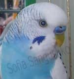
Image 5. Cataract in a budgie (image courtesy Zofia Evangeline Sangushko; used with permission).
Image 6. Cataract in an Amazon (image courtesy Zofia Evangeline Sangushko; used with permission).
Companion birds must be protected from the damaging effects of bright light, both indoor and direct sunlight outdoors. Light coming through windows can damage the eyes, so owners are advised to make sure their birds are in the shade, both indoors and outdoors. UV lamps are an additional cause of cataracts as they shine directly on the bird and can damage the eyes. 22
Birds with cataracts tend to hold their heads to the side so that their good eye is forward. The eyes of older birds should be examined annually to detect early changes in lens opacity (clarity). Because of the small size of the exposed cornea and pupil in psittacines, screening by an avian ophthalmologist is recommended. 37, 10, 60
Many people believe that exposing the bird to direct sunlight or other forms of light, such as lamps directed onto the cage, will not damage the eyes. This is false!
Harrison and McDonald state that, “Exposure to UV light and other direct forms of light will lead to cataracts, macular degeneration, and other forms of age-related eye disease.” 10, 22 They recommend the owner administer antioxidants, such as Vitamins A, C, and E to aid in slowing down the development of these ocular diseases and other age-related illnesses related to exposure to light. 22
Although wild birds will sometimes have eye damage from exposure to bright sunlight, most of the time they find shelter from it in trees and other shady places, so the time they spend in direct sunlight is limited. Between that and the action of the nictitating membrane, their eyes are protected from the damaging effects of bright light most of the time. The brightness of the light is as much of a problem as the ultraviolet rays. Because of this, companion birds must also be protected from the damaging effects of bright light, both indoor and outdoor direct sunlight. Light coming through windows can also damage the eyes. Owners are advised to make sure their birds are in the shade, both indoors and outdoors.22

Image 7. Cataract and posterior synechiae (adhesions in the eye) in a thirteen-year-old canary. Wrinkling of the lid margins is also evident (image courtesy R. Korbel). 33
Cataracts often develop secondary to infection or trauma or may be age-related; they are seen as lens opacities. “If found early in their development, they may be limited to the swelling of the lens fibers, but advanced cataracts involve the entire lens. Eventually, they progress to phacolytic uveitis (breakdown and inflammation of the iris and lens), and blindness results.10, 37, 60
In large psittacine birds, surgical removal of cataracts is successful in many cases. The bird’s general health and the degree to which the cataracts affect its quality of life should be evaluated before surgery. The home environment must be altered for any bird with decreased vision. Early cataracts, especially if uveitis is present, may be painful. NSAIDs, either in as ocular drops or systemic medications (meloxicam, celecoxib) or both, can be used to reduce inflammation and pain. 37
6.1.3 Additional degenerative ophthalmic conditions:
- Keratoconjunctivitis sicca (dry eye syndrome)
- Corneal ulcerations
- Nictitating membrane abnormalities
- Conjunctival granulomas
- Lymphoma
- Lid laxity
- Iris atrophy, leading to darkening of the normally light-colored iris. Sometimes, when the iris atrophies, pupil constriction is seen, causing light sensitivity and retinal damage
- Nuclear sclerosis of the lenses. The nucleus of the lens becomes dense, and fiber production and compression occur.
- Retinal degeneration, often the result of nutritional, congenital, traumatic, or viral conditions. Genetic retinal disease is seen in commercial poultry flocks but not in psittacines.
- Blindness, as a result of pituitary tumors, usually adenomas.
- Blindness, following an acute neurological episode that causes impairment of the Central Nervous System. These episodes are a consequence of atherosclerosis and atherothrombotic stroke (a stroke which occurs when an artery in the brain becomes blocked because of a blood clot or fatty deposits in the blood).
- Glaucoma, usually found in the larger raptor species. It is difficult to diagnose in psittacines because of the small size of the cornea. 6, 37
6.2 Hearing
The sense of hearing in psittacines is neither better nor worse than in humans, just different. Birds can distinguish some ranges of frequencies more accurately than humans can. “These vocalization ranges are comparable to the sounds made by their own species; however, they do not differentiate between the intensity and volume of sounds as well as humans do.” 37 Birds’ hearing loss appears to be less related to aging and more related to loss of function of their hair cells (sensory receptors of both the auditory system and the vestibular system in the ears of all vertebrates); these are able to regenerate. 37
6.3 Olfaction
In the past, it was believed that birds possessed very poor senses of smell and taste. This was attributed to their need to eat quickly due to the danger of predation. But studies have shown that there is a higher percentage of functional olfactory receptors in many species of birds compared to mammals. Early research has shown that some psittacine species have a more highly developed sense of smell than was previously thought. It is not known whether the senses of smell and taste decrease in birds as they age; however, if this is the case, the geriatric bird could be limited in his ability to recognize food, people, and other birds. 37
Images 8, 9. Flaking beak on an 80-year-old blue-fronted Amazon (image courtesy Brian Nadon; used with permission).
6.4 Integument
There is anecdotal evidence that damage to the beak can affect feather grooming and thus affect the appearance of the feather coat. Cutaneous tumors are the most common skin lesions as noted in the ZEPS survey in the table above; these were associated with a history of self-trauma, although the underlying causes was not always evident. 54
According to Reavill and Dorenstein, older lovebirds (over the age of 6) “tend to develop a high number of inflammatory skin lesions (22.8%).” 54 These are primarily the nonspecific syndromes of lovebird dermatitis or chronic ulcerative dermatitis. The most commonly affected areas were the patagium, neck, and back. These areas will develop pruritus, and this leads to the self-mutilation. Although researchers suspect a viral etiology, it has not be identified. Polyfolliculitis may be a part of these lesions. Both syndromes are chronic and often return. 54
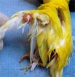
Image 10. Large feather cysts in a canary (image courtesy Julie Burge; used with permission). 
Image 11. Feather cyst in a cockatiel. The blackened area to its left is the wound healing from a previous feather cyst. The second cyst developed immediately after the first was removed from this 22-years old bird (image courtesy Claudia Cano; used with permission).
7. Endocrine System Diseases
Endocrine diseases in older birds are not common. T. Lightfoot states that African grey parrots develop a moderate number of neoplasms, and these often cause sudden deaths. Pancreatic hyperplasia is often found at the same time as hyperglycemia in psittacines. Hyperglycemia is often seen in older, obese psittacines, and it is the cause of avian diabetes mellitus. “Hypertrophy of the parathyroid glands may be seen in birds whose diets are chronically insufficient in calcium. These birds will develop bony lesions which may be seen at necropsy.” 37
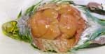
Image 12. Obesity in a budgie (image courtesy Julie Burge; used with permission).

Image 13. Obesity in an Amazon, leading to hepatic lipidosis (image courtesy Julie Burge; used with permission).
7.1 Diabetes Mellitus
According to K. Joyner, diabetes is more common in psittacines than one might think. Glucagon, not insulin, is the regulatory hormone in psittacines. Senior birds, especially females, and usually cockatiels, frequently fall ill to this disease. “Signs include polyuria and polydipsia, which are linked to pancreatic dysfunction. Some birds develop diabetes after having egg-yolk peritonitis, which explains why cockatiels are found with it so often; they have the misfortune of being chronic egg layers.” 32 The inflammatory response to this extends into the pancreas, resulting in damage to the organ. Some birds respond to medical therapy; however, many eventually die from the condition. 32
7.2 Thyroid Disease: Thyroid Hyperplasia or Dysplasia (Goiter)
The presence of goiter in birds is due to thyroid enlargement, and it is most often seen in older birds, particularly pigeons and chickens. It has been found in psittacines as well, mostly budgies. Iodine-deficient foods, usually from all-seed, poor-quality diets, are the cause. “In budgerigars with goiter, clinical changes are limited to regurgitation and dyspnea caused by gland pressure on the trachea and esophagus. The glands can swell to five times their normal size and cause circulatory problems due to compression of the heart and great vessels.” 39 Medication will improve the condition.

Image 14: “A mature budgerigar was presented for a swelling in the thoracic inlet area. The crop was severely distended with food, and the bird had an audible click when it inhaled. Goiter was the presumptive diagnosis, and the bird responded to iodine therapy” (image courtesy E. Hillyer).
It has been known for many years that budgerigars and parakeets (Melopsittacus undulatus) develop goiters when they have been on seed diets which are, by their nature, low in iodine. The birds may appear to only be obese, but other signs may be seen as well. These include:
- Regurgitation due to crop-mucous accumulation and resulting thick mucus that accumulates on the feathers of the head
- The thyroid gland can swell to five times its normal size and cause circulatory problems due to compression of the heart and great vessels.
- Dyspnea (difficulty breathing), caused by gland pressure on the trachea and esophagus. The thyroid gland can swell to five times their normal size and cause circulatory problems due to compression of the heart and great vessels.
- Respiratory sounds such as squeaking from pressure on the syrinx by the swollen thyroid, and dyspnea. 39
Diagnosis is usually easy to make, and the disease is treatable. Dietary changes, the addition of iodine to the water or seeds, and the use of dexamethasone to speed up the response to injectable iodine by decreasing thyroid swelling are the usual treatments for geriatric birds. This treatment has saved the lives of many birds with severe dyspnea. 22, 39

Image 15. Budgerigar (Melopsittacus undulatus). “Severe bilateral thyroid gland hyperplasia, more pronounced on the left thyroid gland. The weight of the left thyroid gland of this bird was equivalent to 6.3% of total body weight (reference value: 0.2%). Bar = 1 cm” (image courtesy Journal of Veterinary Diagnostic Investigation).
8 Respiratory System Disease
As psittacines age, ailments found in the respiratory system are mainly related to repeated insults and inflammatory responses to the insults. Some of these include:
- Dust inhalation
- Irritating gasses
- Microorganisms, such as viruses, bacteria, and fungal spores. 4,15
Continued damage to the lungs and air sacs eventually leads to the development of fibrosis and granulomas. Fibrosis and edema are also caused by circulatory and cardiac disease within the respiratory system. 15
8.1 Chronic Pulmonary Interstitial Fibrosis
Chronic pulmonary interstitial (within the organ’s tissues) fibrosis is being seen in a number of older psittacines in Europe and the U.S. Pulmonary fibrosis causes lungs to become scarred over time. “The tissue becomes thick, and blood does not receive sufficient oxygen. It may occur when an injury to the lungs triggers an abnormal healing response. Toxic substances, allergy, or viral infections may also cause this disease.” 4, 16 It is seen more often in Amazons, and the main symptom is exercise intolerance. Pathologic results show loss of function in the lung tissue, pulmonary interstitial fibrosis, and right-heart failure. 4, 16
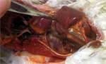
Image16. Cockatoo with congestive heart failure (image courtesy Julie Burge; used with permission).
8.2 Rhinoliths
These are solid concretions or formations of debris that build up in the nares (nostrils). They can result from malnutrition and subsequent squamous metaplasia and chronic respiratory infections. They are also the result of failure to clean out the nostrils when it is needed. As debris builds up, erosions of the nares and operculum (flaps within the nares) take place, resulting in permanent disfigurement of the nares.


Image 17. Rhinoliths as shown in this lovebird. (Image courtesy H. Bowles) 8
Image 18. An old female budgerigar with chronic rhinorrhea and secondary rhinal infection with yeast and/or bacteria. There is discharge from the nares which has accumulated in the frontal
feathers. The cere is dry and appears to have fungal growth around the right naris. It was presented for panting (image courtesy Doneley, Harrison, Lightfoot).16
8.3 Aspergillus
Aspergillus spores are everywhere, and infections often occur when the bird is stressed, suffering from a concurrent sickness, or has undergone trauma, all of which suppress its immune system. Healthy birds exposed to the fungal spores are generally resistant to infections, while immunocompromised hosts exposed to small concentrations of spores are frequently infected. 15
Aspergillosis is one of the frequently occurring mycotic diseases in birds. It is caused by infection from the aspergillus spores. “A. fumigatus is the predominant species of this airborne infection. The spores develop in areas in which the environments are warm and humid and where there is insufficient ventilation and sanitation. They can also come from improperly stored seeds and other feeds. Air sacs are particularly vulnerable because they are warm and oxygenated—a perfect breeding ground for the spores.” 15 R. Dahlhausen states:
“Aspergillus presents as chronic rhinitis and sinusitis, sometimes accompanied by malformation of the nares, beak, and cere, and a purulent nasal discharge. Wheezing sounds may be caused by the formation of rhinoliths or oronasal granulomas obstructing the airways.” 14 A culture is needed to confirm the diagnosis. Most birds present with the chronic form of the disease. Older birds that have been in captivity a long time are most vulnerable to it since the disease is brought on by long-term malnutrition and stress. Older birds are already immunosuppressed, and this contributes to their vulnerability to the disease. 13 Other factors include past disease and overuse of antibiotic or corticosteroid therapy. The chronic form is often seen in African grey parrots, pionus parrots, and Amazon parrots. 13

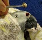
Image 19. Impacted nares from rhinoliths in an African grey (image courtesy Julie Burge; used with permission).
Image 20. Rhinolith removal in the same grey (image courtesy Julie Burge; used with permission).
Bob Doneley, states that, “Older and larger birds (African greys, macaws and cockatoos) often suffer from chronic malnutrition with accompanying Vitamin-A deficiency. This leads to squamous metaplasia and a respiratory environment conducive to Aspergillus spp. as well as propagation and granulomas of the trachea and/or syrinx.” 16 These birds have a guarded prognosis since the disease has gone on for so long that it becomes systemic. 16
Image 21. Granuloma (arrow) of the syrinx in a blue-headed pionus parrot (Pionus menstruus). The bird displayed the characteristic change and weakness in vocalizations several days before it died 13 (image courtesy R. Dahlhausen; used with permission). Copyright © February, 2016. All rights reserved. Images may not be reproduced or used without the express written consent of the owner.

Image 22. Aspergillus fumigatus granuloma exhibiting conidiophore (a type of fungus) spore growth in the lung and air sacs of an African grey parrot (Psittacus erithacus). 13 (image courtesy R. Dahlhausen; used with permission). Copyright © February, 2016. All rights reserved. Images and videos may not be reproduced or used without the express written consent of the owner.
Image 23. Aspergillosis spores in the lungs of a bird (image courtesy Michigan.gov).
9 Renal Disease
9.1 Renal Insufficiency
Kidney disease may be seen at any age, but older birds are more likely to develop renal insufficiency. It is found in a high percentage of birds at necropsy, but there are few non-invasive tests available for it antemortem. Uric acid buildup can be responsible for renal disease, but poor diet or contamination can also cause it. Even if the uric acid levels are normal, the bird may still have renal functional impairment. 58
According to D. Styles, older birds whose diets have been poor their whole lives may display polyuria and polydipsia when placed on a balanced diet. They may also exhibit general malaise and have an increase in their plasma uric acid levels. This may be caused by the chronic deficiency in Vitamin A and the Essential Fatty Acids (Omega- 3 and -6) in their previous diet. This condition is most common in cockatiels. These birds may already have compromised renal function caused by malnutrition, and their urinary production and flow have been obstructed or slowed. “Increased protein levels present in an improved (pelleted) diet may overload their renal and hepatic capacities; consequently, the owner should be very cautious when improving the diet of geriatric birds, and he should make the changes gradually.” 58 Pelleted diets are not advised for the geriatric bird. Owner and veterinarian should determine together how much of a change should be made and when. Vitamins and Omega-3 and -6 fatty acid supplements should be added to the bird’s diet. 58
Image 24. Renal cyst in a cockatiel. The dark green area on the left is part of the intestine; the oval organ above that is the ventriculus, and the tube-like organ above that is the proventriculus. All of the organs have been displaced due to the size of the cyst (image courtesy Julie Burge; used with permission).
Image 25. Renal tumor on a conure (image courtesy Julie Burge; used with permission).
9.2 Gout
Gout is the accumulation of uric acid crystals (tophi) in the articular joints or viscera (internal organs). Articular gout appears most commonly as subcutaneous pockets of a crystalline uric acid with a paste-like consistence in the joints of the feet and legs.” 58 Visceral gout is even more serious and more difficult to diagnose than articular gout. “It appears as whitish accumulations of uric acid in the muscularis layer of vital organs, such as the heart and proventriculus.” 58 It may be seen as a light coating on the tissue surfaces. Articular gout produces a cream- to-yellow-colored deposit in affected joints. 11 The causes include excesses in Vitamin D3, calcium, or protein in the diet; these initiate the renal damage that often leads to gout. 58
Clinical signs for both visceral and articular gout include weight loss, depression, polyuria, and polydipsia. Blood tests and renal biopsy are used to diagnosis the disease, and persistent hyperuricemia (too much uric acid in the blood) is usually present. Radiographs or CAT scans may show small or enlarged kidneys with or without mineralization. Sometimes urethroliths* are seen. Calcification of vital organs often follows visceral gout. Changing the diet from high protein (pelleted) to a diet of seeds and vegetables may help to alleviate or limit the condition over time.41, 58
*A calcification or stone in the urinary passages. Some birds sit very tight on their eggs and fail to void their cloacal contents regularly. Urates can precipitate in the cloaca and lead to the formation of a urolith. Urolithiasis – the presence of calculi, or uroliths, in the urinary passages.
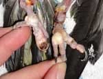
Image 26. Articular gout in a Nanday conure (image courtesy Julie Burge; used with permission).
Articular gout is common in older birds, and it is critical to differentiate between arthritis and articular gout due to the vast differences in progression, quality of life, and prognosis. There are many causes of gout, but the following are the most frequently seen in geriatric birds:
- Glomerulonephropathies (malfunctioning kidneys)
- Renal tubular gout
- Chronic bacterial nephritis (inflammation or infection of the kidneys) 37, 41


Image 27. Gout tophi consist of a painful collection of uric acid crystals in the joints and subcutaneous areas of the feet. (Courtesy B. Doneley, G. Harrison, T. Lightfoot) 16
Image 28. An older budgie suffering from non-specific kidney disease has developed secondary gout deposits in the joints of the lower leg and foot (image courtesy S. Echols) 17
Treatment should involve giving the affected bird supportive care, such as fluid therapy and antibiotics as needed, based on the diagnosis. Once the bird is stable, its diet should be addressed and improved, and the Essential Fatty Acids in Vetomega© (Omega-3 and -6) should be added to help manage the disease. Treatment of articular gout is not always successful; pain relief may be needed, but “NSAIDS for these birds should be given cautiously since they may cause further renal compromise.” 37 If the uncontrolled articular gout causes the bird extreme pain, and the pain cannot be managed, the clinician should discuss euthanasia with the client. 37

Image 29. Visceral gout on the heart and over internal organs of one of the author’s birds (image courtesy Bob Dahlhausen; used with permission). Copyright © February, 2016. All rights reserved. Images and videos may not be reproduced or used without the express written consent of the owner.
 .
.
Image 30. Visceral gout in the internal organs (image courtesy S. Echols).
Image 31. Visceral gout in a Lory (image courtesy Julie Burge; used with permission).
10. Geriatric Conditions of the Integument
Older birds, particularly lovebirds, tend to develop a high number of inflammatory skin lesions. These are mostly dermatitis or chronic ulcerative dermatitis. The most commonly affected areas are the patagium (wing web), neck, and back. These areas will develop pruritis (itching, red, sore areas), and this leads to the self-mutilation. Lesions may be caused by viruses, and Polyfolliculitis may be involved. 54
10.1 Hyperkeratosis
Hyperkeratosis, or abnormal thickening of the outer layer of skin, is common in canaries and is sometimes confused with gout. It may be caused by genetics, particularly in the soft feather species, old age, malnutrition, and hormonal imbalances. Treatment to soften and remove the hyperkeratotic lesions with water-soluble creams is recommended, along with improving nutrition and removing leg bands. 56 Leg band removal is always recommended.

Image 32. Hyperkeratosis is shown in the feet of a canary (image courtesy P. Coutteel)
Clinical signs of hyperkeratosis involving the integumentary system can manifest as overgrowth of the beak and nails, which retain their outer covering due to a proliferation of basal cells. The keratinized outer coatings of pinfeathers are thicker, less flexible and are retained much
longer than normal. Retained coatings prevent pinfeathers from opening, and such feathers appear to be painful to the birds if the unopened feathers are manipulated. Clients commonly report that birds with chronically retained pin feathers are irritable and vocalize as if in pain during preening. While hyperkeratosis is generally associated with dietary deficiencies of vitamin A, excesses of vitamin A are also correlated with hyperkeratosis. The percent of squamous cells present in nasal flushes has been used as an indicator of vitamin A toxicosis. It is important to obtain a full dietary history before prescribing vitamin A supplementation to treat hyperkeratosis. Therefore, a mixture of both vitamin E and vitamin A may be required to treat hyperkeratosis due to a vitamin A deficiency. Deficiencies of zinc and biotin have been associated with hyperkeratosis. Biotin deficiencies, which can result from an excess of salt, are correlated with hyperkeratosis on the footpad and the plantar surfaces of the toes. 56
10.2 Feather Cysts
Although feather cysts are not usually cancerous, they are a type of neoplasm caused by the inability of a growing feather within the follicle to push out to the surface. The feather remains inside the follicle, curled up under the skin. As the feather grows, the lump — caused by the ingrown feather — also continues to grow until the feather cyst becomes an oval or long swelling. At times, it can involve one or more feather follicles at a time.

Image 33. Feather cyst on a parakeet (image courtesy Budgiopolis.word).
A feather cyst can occur anywhere on the bird’s body. In parrots, however, it is commonly seen in the primary feathers of the wing. And although any bird can suffer from feather cysts, it usually occurs in parrots, macaws (blue and gold), and canaries, which usually have multiple feather cysts. The cyst may be opened by the clinician and the ingrown feather removed. Sometimes the follicle must also be removed.
In most birds, feather cysts are caused by an infection or an injury to the feather follicle. In canaries, feather cysts are due to genetic predisposition. (Pet Med MD Feather Cysts in Birds. https://www.petmd.com/bird/conditions/skin/c_bd_Feather_Cysts)

Image 34. A feather cyst (arrow) is shown in a canary (image courtesy Sandmeier and Coutteel). 56
11 Neoplasms and Oncology: Tumors, Masses and Growths
A neoplasm is a new, abnormal growth of tissue which develops as a result of rapid cellular growth. It may be benign or malignant, and it has no physiological function. Budgerigars tend to develop neoplasms as they age at a higher rate than other psittacines. Neurological signs, such as paralysis or paresis (partial paralysis) of the legs, may be evident, and the bird may not be able to maintain its normal position on the perch. Often these growths arise from the liver, kidney, and gonads. 34, 58
Cockatiels and cockatoos tend to form pulmonary carcinomas as they age. Clinical signs depend on where the tumor is found and how large it has become. It is not known what causes these neoplasms. Unfortunately, it doesn’t have a good prognosis. 34, 58
Age influences the development of tumors in avian tissue due to “an increase in genetic damage from environmental agents and errors in gene repair.” 54 The following tumors are often seen in geriatric patients:
11.1 Lipomas
Lipomas are non-malignant, fatty tumors that are most often seen in budgerigars, but Amazons, cockatiels, and other species develop them as well. They are associated with excessive body fat and are usually located on the keel or in the sternopubic (lower abdominal) area. 38
FREE Training to Stop Your Bird's Biting
Get FREE access to Beak School – the only system designed to train your bird. Stop behavioral problems like biting, screaming, feather plucking.
Get the Free TrainingObesity, advanced age, and high-energy diets contribute to lipomas and liposarcomas, according to K. Latimer. In psittacines of any age, xanthomas and liposarcomas may become life-threatening, but they are more quickly fatal in older birds. “Abdominal hernias often develop, and when these are combined with the extensive mass of the lipoma, the bird will have difficulty in evacuating the cloaca, which leads to abrasion, hemorrhage, and infection.” 34 Treatment involves weight loss, improving the environment so that the bird does not traumatize the area, and possibly surgery if all else fails. Complications for the older bird may include hepatic lipidosis (fatty liver disease), decreased liver function, and cardiovascular disease. Any surgery should be as non-invasive and as short a duration as possible. 34
Image 35. Lipoma in a budgerigar (image courtesy Melbourne Bird Vet; used with permission).
Image 36. Malignant tumors in a macaw (image courtesy Oneta Carter; used with permission.)

Image 37. Mesenteric lipoma in a racing pigeon (image courtesy Melbourne Bird Vet; used with permission).
Image 38. Cockatiel tumor (image courtesy Julie Burge; used with permission).
11.2 Air Sac Carcinomas
Air sac carcinomas are rare growths, and it is often difficult to identify the air sac as the tissue in which they originate. They usually occur in older, larger psittacines, such as cockatoos, African greys, macaws, and Amazons. The birds are often brought in initially with cystic masses or bony lesions on the humerus or upper wing bone. 54
11.3 Hemangiomas
Hemangiomas are non-cancerous tumors of the vascular endothelium (a single layer of thin cells that lines the inside surface of blood vessels). They are most often found in budgerigars and are usually seen in the skin of the feet, inguinal region, wing, cloaca, spleen, and the side of the neck. Most birds will develop these when they are around ten years of age. 54

Image 39. Squamous cell carcinoma of the rhamphotheca (entire beak area and bone), and papillomatosis (an abnormal condition of nipple-like growths) in an older Timneh African grey parrot (image courtesy T. Lightfoot). 38
11.4 Hemangiosarcomas
Hemangiosarcoma is the cancerous form of the hemangioma. They usually appear on the beak, wings, feet, legs, and cloaca, and are most often seen in cockatiels. But chickens, swans, Amazons, lovebirds, African greys, pionus, budgies/parakeets, canaries, and finches also develop them. The skin tumors appear inflamed and necrotic. They occur at the same rate in both sexes and around the same age as the hemangiomas. 54
11.5 Hemangiolipomas
Hemangiolipomas, benign tumors of fatty tissue and blood vessels, are not often seen. They are found in the subcutaneous tissue on the body or limbs. In J. Samour’s practice, these have been found on a budgie, a yellow-collared macaw, a cockatiel, a lovebird, an Amazon, and a canary. All of these birds were over 9 years of age. 54
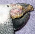
Image 40. Squamous cell carcinoma in the rhampotheca (upper beak) of an African grey parrot (image courtesy Julie Burge; used with permission).
11.6 Hematomas
A hematoma is a collection of blood outside the blood vessel. Causes of their formation include trauma, brain injury, diseases, and infections. Hematomas form when the blood does not clot properly as a result of Vitamin K deficiency. Senior birds may lose their balance and suffer falls or impacts which result in hematomas. Also, as birds age, their skin becomes more fragile. To see the complete series of images dealing with this bird’s surgery, click on the picture to follow the link (image courtesy Peter Wilson, Currumbin Valley Birds, Reptiles, and Exotics Vets; used with permission).
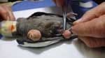
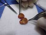
Image 41. Dr. Wilson prepares the leg for surgery.
Image 42. The lump was an encapsulated hematoma, the result of a hemorrhage forming a “blood blister” on the leg. To view, click on figure.
11.7 Neoplasias in specific species
In this table you will find a list of the most common types of neoplasias found in the tissues of various species. The most common tumor of captive parrots overall is the lipoma. 21, 54 Any term that includes the words, “carcinoma” or “sarcoma” refers to cancerous tumors.
Types of Neoplasias
TISSUE NEOPLASIA
|
Muscle |
Leiomyosarcoma (cancer of the smooth muscle tissue, rare) |
|
Bone |
Osteosarcoma (bone cancer) |
|
Kidney |
Renal carcinoma/adenocarcinoma |
|
Testicular |
Sertoli-cell tumor/seminoma (testicular cancer) |
|
Ovarian |
Granulosa cell tumor (cancer of the lining of the ovary) |
|
Proventricular |
Proventricular carcinoma (cancer of the proventriculus) |
|
Hepatic |
Bile duct carcinoma |
|
Endocrine |
Pituitary adenoma (a glandular tumor) |
|
Hemolymphatic |
Lymphoid neoplasia |
|
Fat |
Lipoma |
(Table courtesy Reavell, Dorrenstein, Greenacre) 54, 21
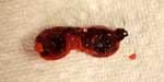
Image 43. Malignant tumor in one of the author’s cockatiels (image courtesy Bob Dahlhausen; used with permission.) Copyright © February, 2016. All rights reserved. Images and videos may not be reproduced or used without the express written consent of the owner. To view, click on figure.
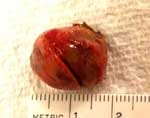
Image 44. Close-up image of the malignant tumor in Image 43 (image courtesy Bob Dahlhausen; used with permission). Copyright © February, 2016. All rights reserved. Images and videos may not be reproduced or used without the express written consent of the owner. To view, click on figure.

Image 45. Necropsy photo of a senior cockatiel hen which presented with depression and severe abdominal distention. Multiple masses were identified in the pancreas and dorsal body wall. Histopathology indicated a pancreatic adenocarcinoma. The cancer had spread throughout the abdominal cavity (image courtesy C. Greenacre) 21
11.8 Facts about Tumors in Birds:
The following information is in CB Greenacre’s paper, “Birds of a Certain Age.” 20
- Over 90% of budgerigars over five years of age will eventually develop leg lameness associated with a renal tumor (usually renal adenocarcinoma) pressing on the sciatic nerve. This is the highest tumor rate of any animal. There is no surgical treatment for this type of tumor.
- Male budgies over five years of age can develop Sertoli-cell tumors or seminomas that secrete estrogen and cause the male’s blue cere to turn brown and produce extra tissue. This is called “brown hypertrophy of the cere.” It can be moistened with warm compresses and peeled away. There is no surgical treatment for this condition.
- Cockatoos and Amazon parrots tend to develop adenocarcinomas more often than any other species. Respiratory adenocarcinomas can be found in the wing, axillary, or lung areas of the birds. The lining of the pneumatic humerus can also develop an adenocarcinoma. In Amazons, bile duct carcinoma is found in the liver, especially if the bird also has papillomatous masses in the cloaca due to psittacine herpesvirus. Surgical removal with wing amputation is the usual treatment method.
- Squamous-cell carcinoma (SCC) is most often seen in Amazons, macaws, and African greys. It is usually found in the mouth or beak area of the Amazons and macaws, whereas the uropygial gland is the disease site most frequently seen in African grey parrots. They may occur anywhere on the body, but are usually found in the oral cavity, the sinuses, on the distal wing, the feet, and the uropygial gland. They tend to be highly aggressive and invasive, and complete excision is usually not possible. Experimental treatment with radiation therapy had being done with some success; however, the tumors are very resistant to this treatment, and long-term control is not generally achieved. Distant metastasis rarely occurs. SCC seems to occur with greater frequency in geriatric birds. The carcinomas create constant necrosis, and the therapies often make it worse, providing a breeding ground for bacteria, yeast, and fungal spores. Throughout any treatment, antimicrobial therapy is advised to avoid septicemia.
- Melanomas are not often seen in birds. They are a type of tumor which spreads throughout the body. These malignant tumors are usually found on the beak, in the liver, on the skin of the face (of birds whose faces are featherless, such as macaws), and in the oral cavity of psittacines. They are known to metastasize to the cardiac muscle, kidneys, brain, and the sinuses.
- Fibrosarcomas can be found anywhere on the body, but are usually seen on the face, in the oral cavity, in the long bones, or in the abdominal cavity. “They are locally invasive and will recur if enough tissue isn’t taken during surgical excision.” The use of radiation therapy is known to control these growths for a long time. Since they do not metastasize well, radiation and/or chemotherapy have been successful with some patients. (These therapies are no longer use since the patients rarely survive them.)
- Internal carcinomas, such as ovarian neoplasias; renal carcinomas; hepatic adenocarcinomas; bile duct, hepatic and pancreatic adenocarcinomas; and carcinomas of the spleen and GI tract have been reported in older birds. “Once as much as possible of the tumors has been removed, and the type of tumor confirmed by histopathology, drug therapy is usually initiated.” 20
Birds tend to have a higher tolerance for some drug therapies than mammals. (R Dahlhausen, personal communication)
11.9 Growths and Tumors of the Reproductive System
11.9.1 Tumors of the Female Reproductive Tract
Senior female birds suffer from condition and tumors of the vent, cloaca, uterus or shell gland, and ovaries. Birds with a long history of egg-laying, egg-binding, and insufficient calcium intake to support these processes will often develop growths and tumors as they age.

Image 46. An elderly, malnourished, female budgie with an obstructive cloacal condition from a uterine tumor. When the obstruction was manipulated, 8 cc of feces were expressed. The owner elected euthanasia (image courtesy B. Doneley, G. Harrison and T. Lightfoot) 16

Image 47. Cystic ovary tumor in a 28-year-old Moluccan cockatoo (image courtesy Reavill and Dorenstein) 54
11.9.2 Tumors of the Male Reproductive Tract
11.9.2.1 Sertoli-cell tumors
Sertoli-cell tumors appear yellow-red and cause enlargement of the testis. The clinical signs are anorexia, lethargy, and dyspnea. They are firm, grayish-white neoplasms (new growths), and the signs are anorexia, dyspnea, cardiac changes, and in budgies, brown hypertrophy of the cere. The average age when diagnosed is 10 years. 34


Images 48 and 49: 6- year-old male budgerigar with a Sertoli-cell tumor (tumor of the ovaries or testes). Note the soft tissue mass in the mid-coelom (main body cavity) and polyostotic hyperostosis (enlargement and thickening of the long bones). Red arrows point to long bones; black arrow to soft tissue mass (image courtesy H. Bowles) 7
11.9.2.2 Brown Hypertrophy of the Cere

Image 50. Brown hypertrophy of the cere and beak overgrowth in a budgerigar (image courtesy Julie Burge; used with permission).


Image 51: A mature male budgerigar with a progressive growth and discoloration of the cere. Brown hypertrophy of the cere is frequently encountered in older budgerigars with gonadal neoplasms. The hypertrophied tissue can be moistened with skin-softening creams and gently peeled away (image courtesy K.S. Latimer). 34
Image 52: Brown hypertrophy of the cere due to hyperestrogenism (too much estrogen) in female budgerigars. Hypovitaminosis A may cause this condition (image courtesy B. Doneley; used with permission). 15
11.10 Tumors of the Uropygial Gland
The uropygial gland has two lobes, and it is located near the base of the tail on the dorsal (top) side of the body. This gland secretes a fatty, pasty, oily, cream-colored sebaceous substance called “sebum.” It is thick in appearance and has a characteristic light, musky smell. The oil is spread on the plumage by the birds during preening to provide waterproofing. The sebum contains the precursors for Vitamin D, and those are converted to Vitamin D when the sebum is exposed to sunlight or artificial UV light. The sebum, now containing the Vitamin D, is then ingested by the bird as it preens. This is the means by which birds absorb Vitamin D. Vitamin supplementation is needed for birds who do not receive sunlight or UV light, or do not have a preen gland. 55
The secretion is carried by a number of ducts to an external papilla, or nipple, covered by a tuft of down feathers forming what is commonly known as the “wick.” One of the most common disorders affecting the uropygial gland is impaction. The gland becomes enlarged, and the wick dries up. Sometimes there is an obstruction with a hardened secretion known as a “lith,” which acts as a plug. Applying warm-water compresses to the area and gently massaging it and then expressing the sebum from the gland usually resolves the obstruction. 55 Sometimes the gland needs the care of an avian veterinarian. 55

Image 53. Adenocarcinoma of the uropygial gland (image courtesy Olanthe Animal Hospital; used with permission)..
Image 54. Uropygial gland tumor in a budgie (image courtesy Julie Burge; used with permission).
When the bird is at the practitioner’s office, its uropygial gland should be examined for evidence of abnormalities, such as enlargement, inflammation, impaction, abscessation, and neoplasia. Any of these may lead to self-mutilation or trauma by the bird. 55
Some species of birds possess a uropygial gland, and others do not. Most psittacines have this gland.
8.10.1 Species which do or do not possess a uropygial gland: 5
Uropygial Gland Absent
Argus pheasant
Citron cockatoo
Woodpecker
Cormorant
Double-headed Amazon
Yellow-Fronted Amazon
Cassowary
Green-winged macaw
African Grey
Sun Conure
Tailless Domestic Fowl
Umbrella Cockatoo
Rock Dove (pigeon)
White carneau and rumples pigeon
Blue and Gold macaw
Uropygial Gland Present
African Grey
Eclectus
Goffin Cockatoo
Budgerigar
Gold-capped conure
Cockatiel
Indian Ringneck
Grey-cheeked parakeet
Muluccan cockatoo
Hyacinth Macaw
Red-masked conure
Red-cheeked conure
Rose-breasted cockatoo
Emu and ostrich
Severe macaw

Image 55. Fibroses developing on the uropygial gland of one of the author’s birds (image courtesy J. Miesle).
Uropygial gland tumors can be either adenomas or carcinomas, and it is difficult to tell the difference visually. Both will appear as swellings and may be inflamed. “Adenomas are usually well circumscribed and encapsulated, whereas carcinomas appear to be less defined and will infiltrate the surrounding tissue.” 57


Image 56: Squamous cell carcinoma of the uropygial or ‘preen’ gland in a sulphur-crested cockatoo (image courtesy Melbourne Bird Vet; used with permission).
(http://www.melbournebirdvet.com/interesting-cases.aspx)
Image 57: Uropygial gland adenocarcinoma (cancer in gland tissue). An older male cockatiel was presented with a several-month history of poor generalized feather condition and feather loss around the uropygial gland. A raised, firm, mass was evident. The mass was surgically removed, and the histopathology confirmed the diagnosis (image courtesy K.S. Latimer). 34
11.11 Xanthomas
Xanthomas are growths that occur usually on the wing but can spread to the body and occur internally, in or around the heart. They are friable, yellow-colored, fatty-appearing masses that may be found anywhere on the body, but the most commonly seen places are on the distal wing and in the keel area. They are found mostly on older psittacines. 46 They are not true neoplasms since they are caused by excess fat and cholesterol in the diet, usually from all-seed diets and lack of exercise. As the fat and cholesterol build up, the xanthoma continues to grow until it is so huge that surgical excision of the mass may be required, and sometimes amputation is necessary. 46 If attended to early in the disease process, the xanthoma may be corrected by dietary changes, exercise, and a non-surgical treatment protocol which the author of this paper has developed. 46
The disease often causes inflammation of underlying tissues. When located at the wing tip, the mass may cause the wing to droop, resulting in trauma to the mass, inability to fly, and an altered gait. The diet should be changed to one that is low in protein and fat. 46 Serum cholesterol levels should be closely monitored; they are usually elevated in birds with xanthomatosis and should be medically reduced to a normal level prior to surgery. 6
The average age of affected birds is 10 years (range 3-30 years), and they are found most often on cockatiels and budgies. They may also be found on lovebirds, Amazons, macaws, cockatoos, and African greys. According to Reavill and Dorenstien, “They are masses of foamy macrophages, giant cells, and cholesterol deposits that produce thickened, dimpled skin with yellow-orange coloration. They may occur in internal organs.” 54 The aortic arch tends to hold onto the cholesterol. They are very vascular, and “may affect the brain where it appears in association with blood vessels.” 5 Because they are so vascular, any surgery performed must be done very carefully as they will bleed easily. Cold-laser therapy is being used on them with some success. 54
There is evidence that ginseng reduces the fat and cholesterol deposits within the xanthomas and within and around the heart. The author’s cockatiel responded favorably to this herbal supplement, recommended by Bob Dahlhausen.

Image 58: A 10-year-old female cockatiel was presented for feather-picking associated with the right carpus of the wing. A diffuse (spread out), firm, yellow mass was noted on physical examination. The xanthoma was surgically excised. (Image courtesy K.S. Latimer) 34
Image 59. The left underwing xanthomas on the author’s rescued cockatiel. Note the necrotic lesions under the wing and xanthomatous tissue on her axial areas—under her wing and on her sides (image courtesy J Miesle). 46
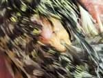
Image 60. The same bird with xanthomas on the left-wing elbow joint. The xanthoma had covered the entire joint, but now all that is left of it is the lower, yellowish, dimpled part where the feather is coming through. The pink area above it is bone and skin tissue. Those feathers eventually fell out as the xanthoma reduced in size (image courtesy J. Miesle) 46
Image 61. An extreme case of diffuse and discrete xanthomatosis. This bird had lived with this for several years before passing away from the damage caused by the xanthoma. This bird had another large xanthoma under the left week and on the back (image courtesy Belinda James; used with permission).
12 Musculoskeletal System
Elderly birds often present with degenerative lesions on their joints. The causes may include a past trauma, infection, or metabolic condition such as gout. The joints are enlarged, and there may be erosion of the cartilage. “Cartilage flaps and free pieces may fill the joint cavity. The tissue eventually forms fibroses in the joint capsules and the soft tissue surrounding them. Muscle-wasting and joint stiffness were reported in macaws over 40 years of age. The range-of-motion was limited, particularly in the intertarsal joints. There may also be twisting deformities that develop in the carpal bones of the wings, causing the primary flight feathers to twist away from the rest of the wing.” 12 (This is sometimes the cause of “Angel Wing” disorder.)
12.1 Arthritis and Bone Disease
Arthritis (inflammation of the joints) often occurs in geriatric birds. The birds suffer from limited range of motion in their joints. The bird’s weight, general physical condition, previous injuries, and any other concurrent medical conditions can all contribute to the onset and severity of arthritis. 24
Birds can develop septic arthritis (infection caused by a pathogen) and traumatic arthritis (caused by an injury) at any age, but senior birds are most prone to acquiring these diseases. Septic arthritis is most often seen developing in the digits. Osteoarthritis is also commonly seen in geriatric birds; it can lead to other issues such as pododermatitis if not discovered early and treated. 24
Clinical signs vary, depending on the location of the arthritis and the severity of the disease. These signs include:
- Lameness or crippling
- Lower level of activity
- Lack of desire to fly or not flying as well as before
- Inability to perch normally or falling off perches
- Swollen or warm joints
- Decreased range-of-motion
- Feather-picking or mutilation
- Excessive vocalization 15,24,37,54
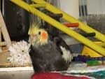
Image 62. An 18-year-old cockatiel, owned by the author, with arthritis. It is typical of birds to find soft places to sit and to sit low instead of perching in an erect position. It is easier on the joints, and the bird will keep his legs warmer with that position. This bird is receiving Procosa®, a glucosamine and chondroitin supplement suspended in Vetomega®, and Celebrex® for discomfort. He sleeps in a bin on towels for comfort instead of in a cage (image courtesy J. Miesle.)


Images 63, 64. Image on the left is of an adult swan which was presented with intermittent lameness. The tibiotarsal joint was hot, firm, and swollen. Radiographs indicated joint enlargement, subchondral bone lysis (breakdown) and erosion of the intercondylar space (the smooth surface area at the end of a bone), forming part of a joint. These lesions were suggestive of septic arthritis. Radiograph of the right is the normal leg for comparison (images courtesy M.C. McMillan).55
The diagnosis of arthritis is based on clinical signs, the physical examination, and radiographs or CT scans. Diagnostic imaging may reveal:
- Narrowing of the joint space
- Sclerosis of subchondral bone (hardening of the bone tissue lying under cartilage)
- Misalignment of the joint
- Osteophyte formation (bony projections that form along the joint margins) 15,24,37,54
CT scans help determine the severity of the bony changes. Commonly affected joints are the foot, knee, and toe joints. The joints of the wings appear to be less frequently affected 24 Treatment is multimodal, with NSAIDS such as Celebrex® normally prescribed.
If the bird does not respond to these and conditions worsen, opioids may be necessary. 15 Additional management includes:
- Changes in perches to softer perches (rope perches, wrapped perches, or padded perches) to reduce stress on weak or painful legs or feet. Vetrap or other cohesive wrap will provide softer perching.
- A weight loss and exercise plan to reduce stress on legs and feet
- A healthier diet (rich in Omega-3 and 6 fatty acids)
- Physical therapy 48
- Use of a large storage container or bin with towels instead of a cage for crippled birds. 49
Flying in a safe environment is the best form of exercise. If that’s not possible, the bird should be encouraged to climb, walk, and step-up multiple times. He may also receive additional exercise by foraging for food in different areas of the cage, floor, or enclosure to encourage movement. 15, 24, 37, 54
According to Bob Doneley, old age is not the only cause of osteoarthritis and the chronic pain that follows. It can also be caused by “trauma, infection, immune-mediated disease, or developmental malformations. Pain develops following joint dysfunction, muscle atrophy, and limb disuse.” 15 The pain varies, depending on the severity of the injury. 15
Studies have shown that obesity is a risk factor for osteoarthritis in many species. Treatment may involve adding foods containing Essential Fatty Acids. Fatty acids have an anti-inflammatory and antioxidant effect and protect the kidneys. Flax-seed oil, an ingredient in Vetomega®, given orally, is recommended as the best source of fatty acid supplementation for birds. 24, 48
12.2 Use of Cold-Laser Therapy in Arthritic Birds
“All species get arthritis. We recently invested in a Class IV Companion laser. We have seen some remarkable changes in a couple of Moluccans that were feather picking at the points of arthritis—one over its back, the other on both its carpal joints. Radiographs confirmed the lesions, and laser therapy has done wonders where pain meds such as Metacam® (meloxicam) have not helped. We’ve also seen some positive results in self-mutilation. Like drugs, it requires ongoing maintenance; but unlike drugs, there are no side effects. The new Companion CTX actually has settings for birds and other exotics. Do not forget aging joints. Everyone worries about the internal damage, as they should be, but we also need to be concerned about joints and external discomfort.” (Bart Huber, personal communication)
12.3 Osteoporosis
Osteoporosis is a metabolic bone disease which involves the mineralization of bones. Birds may exhibit folding fractures and severe bending of long bones.55 Osteoporosis often causes paresis or paralysis of the leg bones and feet, but it is sometimes seen in the wings.53 These birds may have to be euthanized due to excessive pain. 55
Sometimes referred to as “cage-layer fatigue,” it is most often seen in birds (especially chickens) that are confined to a cage and used for egg production. “Immobilization, combined with calcium deficiency, can cause the moderate osteoporosis to deteriorate into severe osteoporosis with advanced clinical signs. Birds are found paralyzed in their enclosures, with skeletal deformities and enlarged parathyroid glands. These are caused by spinal cord compression due to fractures and insufficient calcium.” 39

The main causes of osteoporosis for elderly birds are:
- Chronic egg-laying in reproductively active hens
- Increased phosphorus intake
- Calcium and Vitamin D3 deficiencies
- Reduced physical activity
- Poor diet (mainly all-seed diets), which leads to excessive dietary phosphorus which in turn leads to insufficient calcium absorption. 39
Radiographs and plasma levels of calcium, ionized calcium, and Vitamin D are needed to determine appropriate therapy. 8, 37
“Here are some radiographs of a 50 – 70-year-old Blue Fronted Amazon. It currently belongs to a rescue; they have had it for 4 years, and the age is an estimate from the family that donated her after her owner passed away. More important are the lesions. She presented for falling then drooping her left wing. She has a localized osteoporosis of her left humerus, which is fractured, displaced and very narrow. She also has two fractures of her left ulna which are more recent. The rest of her bones are OK for her age. She is not exhibiting any pain and attempts to use the wing; she is eating, drinking and seems content” (Bart Huber, personal communication).
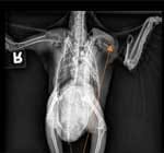
Image 65. Left humerus (image courtesy Bart Huber; used with permission.)
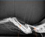
Image 66. Left ulna and left humerus wing (image courtesy Bart Huber; used with permission).
13 Pododermatitis (Bumblefoot)
Pododermatitis is a relatively common condition in older, obese, sedentary pet birds. “It is a general term for any inflammatory or degenerative condition of the avian foot and can range from mild redness to changes in the structure of the foot. Bumblefoot develops most commonly when birds are either housed with inappropriate perching or secondarily to an injury in one leg, which causes the bird to shift its weight to the uninjured leg. This creates increased pressure and potential ulceration on the plantar surface of the foot.” 49
Birds most at risk are those with leg fractures; arthritis of a hip, stifle, or tarsal joint; and with the complications stemming from obesity and poor diet, which lead to Vitamin A deficiency. These are the same conditions that can predispose a bird to arthritis. Pododermatitis is often a sequela of arthritis or osteoarthritis. 14 It is also a result of exposure to tobacco smoke. 49
Pododermatitis can be both the cause and result of decreased activity in the senior bird. Malnutrition plays a key role since it decreases the integrity of the plantar epithelium (skin tissue on the bottom of the feet) and leads to obesity, which puts additional stress on the feet. The cage environment, especially the perches, is important in providing comfort and stability for arthritic birds with bumblefoot. 57 If the bird is lame in one leg, the weight is borne on the other leg, and this can lead to pressure necrosis, infection, and subsequent pododermatitis. Therefore, the other leg should always be carefully examined. 16
Bumblefoot occurs on the plantar surface of the foot and affects the metatarsal and digital pads in particular. Decubital ulcers (ulcers which occur on the skin surface due to prolonged pressure) form, and sometimes staphylococcus infections develop as a result. 57 There may be loss of definition of the epidermis (seen as a shiny, reddened surface), swelling, erosions, ulcers and scabs. Pododermatitis is common in captive raptors, but is never seen in the wild, even in one-legged birds.
Pododermatitis is a progressive disease. A localized hyperemic lesion (an excess of blood in the vessels supplying an organ or other part of the body) develops, followed by ulceration, and if untreated, abscess formation and osteomyelitis. Initially, the skin on the metatarsal and digital pads becomes flattened, shiny, and smooth. The skin may become proliferative and then ulcerate, allowing bacterial access, which leads to inflammation and infection. As the infection progresses, tendon sheaths become affected, and osteomyelitis and septic arthritis develop.16, 48
Lesions are classified from Grades I to VI, Grade VI being the most severe. Birds who have pododermatitis are in danger of losing their dermal tissue and even requiring amputation if the condition is not treated early and changes made in their perching and husbandry. “Older budgerigars and cockatiels (five to ten years old) may have a Grade V to VI lesion if precipitating factors are not corrected early. The disease may progress into bone changes and osteomyelitis. Prognosis for full recovery of Grades I to IV is more favorable than Grade V to VI lesions.” 14, 15, 48
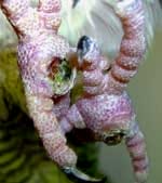
Image 67. Pododermatitis, later-stage lesions in a cockatoo (image courtesy Julie Burge; used with permission).
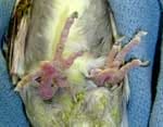
Image 68. Pododermatitis in mid-stage lesions in a cockatiel (image courtesy Julie Burge; used with permission).
Birds may present with lameness, depression, and anorexia due to inflammation, pain, and infection. Diagnosis is based on clinical signs, physical examination findings, radiographs, and culture results. Affected birds should be examined thoroughly for predisposing injuries or illness. Treatment ranges from topical creams for mild cases to surgery and bandaging for extreme cases. Husbandry changes are vital. Birds require correct perches—those that provide variety of diameter, materials, and texture—as these are of utmost importance in preventing and treating pododermatitis. All rough-coated perches, such as concrete, sand-covered perches, dowel perches, heated perches, pumice, and any abrasive material, need to be removed and replaced with padded or rope perches. (Heated perches should never be used because of the burns they can inflict on the feet and the rough coatings on them.) Hard perches should be wrapped with Vetrap or another such cohesive wrap to provide a heavy padding for the bird. Perches that are supposed to keep nails filed do not work but only irritate the skin, leading to pododermatitis. Nails should not be trimmed too short, and wings should not be clipped since clipping affects the bird’s ability to balance. Nutritional deficiencies need to be addressed. 23, 46
14 Cardiovascular System Diseases
14.1 Heart Disease
As birds live longer and diagnostic techniques improve, cardiac disease is being diagnosed more frequently.20 Heart disease in birds with enlarged, poorly functioning hearts can be difficult to detect and may mimic other problems, such as respiratory, hepatic, or ovarian disease. Cardiac disease has been associated with atherosclerosis in pet birds, and potential risk factors are a sedentary lifestyle, a high-fat diet, and too much cholesterol in the blood. 37
Clinical signs are weakness, depression or lethargy, increased respiratory rate and effort, and tachycardia (rapid heartbeat.) With right-sided heart disease, hepatomegaly (enlarged liver) and ascites (fluid in the body cavity) are common. In birds, right-sided cardiac disease is more prevalent than left-sided. Disease also may be subclinical, then flare up suddenly, with the bird going into cardiac arrest when diagnostic tests or treatments are attempted. Diagnosis is based on echocardiogram, and treatment includes the same cardiac drugs used in mammals. 20

Mass on Neck Axillary Mass Calcified Aorta
Image 69: Masses on neck and axilla and calcified aorta (image courtesy Bart Huber; used with permission).
In this image of the bird, “on the lateral view of her body, you can see a white spike, kind of knife-shaped. That is a calcified aorta, arteriosclerosis. We can also see a small mass in her neck. These are geriatric issues. The fracture brought her in, and the heart and mass were incidental findings that will complicate her surgery (amputation) and recovery. There is also a mass in her left axilla (image courtesy Bart Huber; used with permission). 31
Consultation with a cardiologist on any avian patient with suspected cardiac disease is advised. Diagnosis of the cardiovascular abnormality and the formation of a therapeutic plan require knowledge of avian anatomy and physiology as well as a veterinary cardiologist’s diagnostic skills and medication recommendations. Radiographs and cardiac ultrasound can aid in the diagnosis. 37
FREE Training to Stop Your Bird's Biting
Get FREE access to Beak School – the only system designed to train your bird. Stop behavioral problems like biting, screaming, feather plucking.
Get the Free Training“The stress of handling can increase intracardiac blood flow velocity 300% in avian patients; therefore, inhalant anesthesia or sedation is preferred over manual restraint when echocardiograms are performed in all but the most docile birds.” 37
Although most avian therapeutic regimens are still taken from those used in mammals, there are reports that indicate that cardiac drug therapy can improve cardiac function, thereby increasing the quality and length of the bird’s life 37, 43
14.2 Pulmonary Hypertension
The cardiovascular system of birds differs anatomically and physiologically from that of mammals. Mammals are able to maintain sufficient pulmonary vascular resistance, but birds are not. “This results in the inability of the pulmonary vasculature to accommodate increased cardiac output by either altering vessel diameter or changing the number of vascular channels being used. This causes the high incidence of pulmonary hypertension syndrome in poultry and right-sided heart disease in psittacine birds. For geriatric psittacine patients with pulmonary hypertension, vasodilator therapy should be explored. Macaw asthma may cause pulmonary hypertension from chronic capillary hypoxia” (too little oxygen).37 Some chronically obese birds will display infiltration of fat into the myocardium (the muscular tissue of the heart), and this can be seen at necropsy. The bird will then suddenly succumb to the disease with no other findings on histopathologic examination. 43
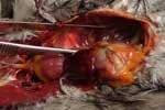
Image 70. Cockatiel with pulmonary congestion (image courtesy Julie Burge; used with permission).
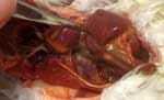
Image 71. Cardiomegaly from pulmonary congestion (image courtesy Julie Burge; used with permission).
14.3 Atherosclerosis
Atherosclerosis is a chronic inflammatory and degenerative disease of the arterial wall. The blood vessels are narrowed due to progressive accumulation of inflammatory cells, fat, cholesterol, calcium, and cellular debris which form fibrous and fatty plaques within the blood vessel. It may lead to stenosis (narrowing of the blood vessels), ischemia (poor blood supply to the heart or brain), thrombosis (blood clot in a blood vessel), hemorrhage, and aneurism (bulge in a blood vessel). Amazons and African greys have the highest numbers reported, followed by cockatiels, lovebirds, Eclectus parrots, cockatoos, and macaws. 37
Atherosclerosis is caused by high-fat, high-cholesterol diets, lack of exercise, age, species susceptibility, and exposure to some infectious agents. The result is long-term, chronic inflammation. The type of dietary fat consumed affects the development of atherosclerosis more than the total amount of fat consumed. 37 Diets high in Omega 3 and 6 fatty acids and herbs protect against atherosclerosis in geriatric parrots.” 47
Atherosclerosis leads to stroke, heart attack, and vascular disease and is seen in parrots with increasing frequency. The clinical signs for birds include circulatory conditions, lethargy, dyspnea, fainting, sudden falling, nervous symptoms due to blood loss in areas of the body, and sudden death.


Image 72. A 32-year-old green-winged macaw was presented for progressive weakness of several weeks’ duration. The bird was recumbent, depressed and severely dyspneic. The bird died shortly after presentation. Necropsy indicated a pale, mottled heart. Histopathologic changes included atherosclerosis and myocardial fibrosis (impairment of the heart muscles) (image courtesy: K. Latimer). 35
Image 73. Necropsy photo of African grey parrot (Psittacus erithacus) with arteriosclerosis. Note the thickening and discoloration of the wall of the large vessels (arrows) (image courtesy M. Pees).50
Atherosclerosis refers to damage from lesions in the arteries. It is being seen more often in geriatric psittacine birds; however, African grey parrots may develop this at a younger age. “Atherosclerotic plaques in the coronary vessels cause abnormal vascular flow and loss of integrity of the heart’s lining. These changes in vessel walls can initiate thrombosis. In birds, lesions are primarily in the aorta and brachiocephalic arteries (a blood vessel that originates from the aortic arch). 37 One study showed that 86% of the Amazon parrots with atherosclerosis who were in aviary and zoo populations were over eight years old, and many were over 15 years old. In one report, the incidence of atherosclerosis in the study group was 91.9% in African greys and 91.4% in Amazons. 37, 43
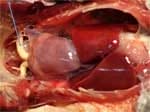
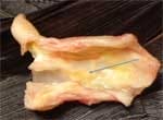
Image 74. Atherosclerosis showing the plaques in an African grey (image courtesy Julie Burge; used with permission).
Image 75. The great vessels of the heart open to show the plaques in the vessel walls (image courtesy Julie Burge; used with permission).
Risk factors include:
- Increased age (most affected birds are 10–15 years old or older)
- Reproductive disease (predominantly female) due to the effects of estrogen on lipid, protein, and calcium metabolism in reproductively active female birds
- Hepatic disease
- Concurrent myocardial fibrosis (abnormal thickening of the heart valves)
- High-calorie, high fat diets
- Hypercholesterolemia (high levels of cholesterol in the blood)
- Lack of exercise, sedentary lifestyle. 18
Clinical signs include:
- Diminished blood flow through the arteries
- Severe lesions in the heart
- Other cardiac disease
- Congestive heart failure
- Sudden death 18
Patients are often presented for:
- Falling or collapsing and exercise intolerance
- Transient or persistent weakness and lethargy
- Dysfunction of one or more limbs
- Altered and reduced mentation, such as disorientation or confusion
- Blindness or anisocoria (unequal size of the pupils)
- Seizures, tremors, or convulsions
- Vestibular proprioceptive signs (perception of body position and movement)
- Paresis of one or both pelvic limbs
- Extension and rigidity of one leg and wing
- Clenching of the toes
- Ataxia and difficulty perching
- Regurgitation
- Dyspnea (difficulty breathing)
- Ascites (fluid build-up in the abdomen) 18
Although it can occur at any time, one sign that is often observed in the practitioner’s office is syncope (a temporary loss of consciousness caused by a drop in the blood pressure) during exertion. 37 Often the client reports sudden symptoms which resolve a short time later. These signs are consistent with stroke, but rarely is this confirmed diagnostically. It can be difficult to distinguish between atherosclerosis and stroke. 18
14.4 Diagnosis and Treatment
Diagnosis can be challenging. “Radiographically, the right aortic arch may be enlarged and show increased density. Lipemia (fat in the blood) is often present, and marked increases in cholesterol and triglyceride concentrations may be seen. Unfortunately, definitive antemortem tests are lacking.” 18
Medical treatment is anecdotal. A variety of treatments have been advocated to lower cholesterol levels, but none appears to be consistent in its efficacy. Essential fatty acids (flax seed oil) have been advocated and used to reduce cholesterol and inflammation. Weight loss, increased activity, and dietary improvement will improve the overall health and hopefully prevent atherosclerotic plaques. In some cases, clinicians may choose to begin therapy with lipid-lowering medications such as statins. Some studies have shown that ginseng is effective is lowering the plasma cholesterol levels in birds. Atherosclerosis, degenerative heart disease, and congestive heart failure should be considered in any geriatric patient with the above signs. 37
At necropsy, grossly thickened arterial walls are seen, as are cholesterol layers within the aorta and large arteries. Microhemorrhage, metaplasia, fibrosis, and mineralization are also found. “These lesions cause increased arterial resistance that affects the heart. Right-heart failure can lead to congestion, atrophy, and eventually cirrhosis of the liver. Sudden death is commonly seen in birds who appear to be in good condition and even overweight. Birds experience decreased blood supply to the brain as a result of severe narrowing of the carotid arteries. He may lose awareness of his surroundings in the days or weeks before his death.” 58
I also see cholesterol deposits in the great vessels of the heart. The great vessel walls should be flexible, but instead they are hardened. It all relates to nutrition—decades of improper nutrition—mainly from all-seed diets.
Dr. Bob Dahlhausen

Image 76. Cockatiel with lung congestion and heart failure (image courtesy Julie Burge; used with permission).


Image 77. Atherosclerosis of the heart. A 20-year-old Rosella was presented with a history of feather dystrophy and exercise intolerance. The bird tested positive for PBFD circovirus and died shortly after presentation. At necropsy the great vessels were noted to be hard, irregularly shaped, and yellow. The vessels were partially calcified, and the histologic diagnosis was atherosclerosis (image courtesy K. Latimer, P. Rakich). 35
Image 78. Necropsy on a blue-fronted Amazon showed pericarditis urica (abnormal amount of fluid between the heart and pericardial sac surrounding the heart). Uric acid deposits can be seen on the pericardium (lining of the heart) that covers the heart and the serosa (enclosing membrane) of the liver. The yellowish discoloration of the large vessels is an indication of arteriosclerosis (image courtesy M. Pees) 50
15 Chronic Liver Disease
Chronic liver disease is the result of repeated damage to the liver over the lifetime of the bird; hence, older birds are more vulnerable to it. Unfortunately for many birds, by the time it has reached its end stage (cirrhosis or fibrosis), it is too late for treatment to be effective. Many birds show no signs until the very end. 27
The possible causes include:
- Toxicities, such as therapeutic agents or naturally occurring toxins
- Chronic cholangitis (obstructive biliary disease, or infection of the bile duct)
- Chronic congestion from right-sided heart failure
- Metabolic disorders, such as hemochromatosis (Iron Storage Disease) or hepatic lipidosis (fatty liver disease)
- Chronic, active hepatitis
- Inflammation of the liver
- Malnutrition 27,44
15.1 Hepatic Lipidosis or Fatty Liver Disease
Liver disease can affect birds of any age, but it is more common in geriatric birds. The most commonly seen liver disease is Fatty Liver Disease. It most often is the result of long-term feeding of a high-fat diet and sedentary lifestyle. Chronic liver disease can result in fibrosis of the liver, decreased liver function, and liver failure. Amazons, cockatiels, macaws, and budgies are highly represented in cases of hepatic lipidosis. 54
Clinical signs of liver disease and liver failure are:
- Anorexia and lethargy
- Enlarged, hardened liver
- Overgrown beak and nails with bruising
- Biliverdinuria, a condition which creates green, unusual droppings
- Discolored and poorly conditioned feathers
- Ascites in later stages
- Glossy, black coloring in the feathers due to the exposure of melanin from total loss of normal green or blue pigment.
- Weight loss and diarrhea
- Bruising or bleeding of the skin and prolonged clotting time
- Abdominal hernia and distension 7, 27


Image 79. Polyurates with biliverdinuria and feces typical of enteritis (image courtesy Doneley, Harrison, Lightfoot) 16
Image 80. Egg-bound pearly cockatiel passes a loose, liquid dropping after having the egg removed. Liver stress is the cause of the condition in this bird. (Notice the darkening of the feathers and greasy appearance.) (Courtesy B. Doneley, G. Harrison, T. Lightfoot) 16
Birds who take in excessive fat and carbohydrates in their diet, are overfed, and are sedentary (particularly Amazons) are predisposed to hepatic lipidosis. 27 They are usually overweight or have a history of being obese. They first begin to show signs when they have become stressed from illness or other issues and are now anorexic and depressed. They have a high accumulation of lipids in the blood and are unable to store glycogen in the normal way. The bird becomes ill and doesn’t eat, and then the glycogen stores are depleted. The bird eats even less and becomes more depressed. This cycle of increasing energy depletion results in death unless there is medical intervention. 27, 42


Heart Liver Fat
Image 81. Severe fatty-liver degeneration and bacterial hepatitis in a 23-year-old Amazon parrot hen with ovarian cysts (image courtesy J. Lumeji, Clinical Avian Medicine, Harrison, 2006). 41
Image 82. Fatty liver in a Mexican red-headed Amazon (image courtesy Harrison and McDonald, Clinical Avian Medicine, 2006). 22
Image 83. The marked change in color and liver texture is characteristic of cirrhosis (image courtesy Hochleithner and Hochleithner).27
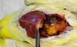
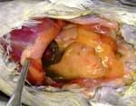
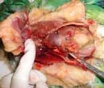
Images 84, 85, 86. Liver abscess in a cockatiel image courtesy Julie Burge; used with permission). $
- I
- High triglyceride and cholesterol concentrations.
“Liver biopsy may be necessary to confirm the diagnosis and determine etiology and prognosis. The affected liver is visibly shrunken, pale and fibrotic. The capsule is thickened, and the edges are rounded.” 30
16 Bacterial diseases
Senior birds are as vulnerable to bacterial diseases as birds of younger ages, but there is one bacterium that appears to strike them more often: Salmonella.
16.1 Salmonella
People with healthy immune systems can generally fight off the salmonella pathogen, but if they are carrying the bacteria, they can infect their companion birds since this is a zoonotic disease. Geriatric birds are more susceptible to this disease, and human-to-animal interactions have been shown to be a cause. 40 Species which are most susceptible are African grey parrots, Amazon parrots, cockatoos, and macaws. Contact with reptiles often leads to the development of this disease in birds. 40 Infections can lead to septicemia and death. It is spread generally through the oral route, although it can become airborne and enter the body in that way. It is also spread vertically, from the hen to the egg. The chicks hatch and spread it throughout the nursery. Vertical infection can also occur when the hen feeds the chicks with contaminated crop contents. According to Helga Gerloch, “Signs include lethargy, anorexia, polydipsia (sometimes followed by polyuria), and diarrhea. In subacute to chronic cases, CNS signs, arthritis (particularly in pigeons), and dyspnea occur. Indications of liver, spleen, kidney or heart damage are common. With high-dose infections, conjunctivitis and other eye infections may occur.” 19
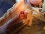
FREE Training to Stop Your Bird's Biting
Get FREE access to Beak School – the only system designed to train your bird. Stop behavioral problems like biting, screaming, feather plucking.
Get the Free TrainingImage 87. Streptococcus infection on the patagium of one of the author’s elderly female cockatiels (image courtesy J. Miesle).
16.2 Other Bacterial Diseases
Bart Huber discusses the bacterial diseases he sees in his practice among older birds: “I rarely see salmonella, and we culture a fair number of birds, probably 8-10 a week. These are birds of all ages as it is part of our preventative panels as well as working with ill patients. Most commonly we find E. coli, Enterobacter sp., Klebsiella and Pseudomonas aeruginosa and other Pseudomonas sp. Quite frequently these are asymptomatic. We have seen a rise in Methicillin Resistant Staphylococcus Aureus, S. pseudintermedius and MRSP and MRSSp.
“Of all these, the most challenging to get rid of tend to be the Pseudomonads. I come from the school of thought that psittacines should not have gram negative bacteria, so I do tend to treat it unless it’s a light growth and very susceptible. Then I will focus on diet. cleaner environment and probiotics.
Older birds, especially African grays and cockatoos, are at a higher risk of aspergillus and other fungal diseases.”
17 Reproductive Disorders
The reproductive lifespan of birds varies according to the species. Some Congo African greys are still producing at 40 and laying infertile eggs at 60 years of age. Greg Burkett reports an African grey which laid its first egg at over 20 years old. He has also had some cockatiels over 20, greys over 25, a macaw over 30, and an Amazon over 50 who have all laid eggs. 10 Dr. Dahlhausen has had in his practice a scarlet macaw that laid her first egg at the age of 95! (R. Dahlhausen, personal communication.)
There have been many studies of the effect of aging on reproduction, primarily in domestic poultry. “There is a definite decline in fertility and reproductive/hormonal behavior as the bird ages, and clutches decrease in number and frequency. There is also a decrease in the viability of the young. The ovulatory cycle and spermatogenesis (sperm production) both cease.” 10, 62 In a study of macaws in Parrot Jungle and Gardens in Florida, the oldest birds to successfully breed were 35 years old; but generally, reproductive activity declines in the twenties to thirties. Dr. Burkett states that budgies are able to reproduce much later in their lifespan compared to other species, and most birds live at least one-third or more of their total lifespans after reproduction ceases 10,62.
17.1 Infertility
Older breeders tend to become obese, and that is one of the primary factors in infertility. They also tend to suffer from the effects of:
- Inbreeding
- Damaged vent feathers
- Drug therapy, causing vitamin deficiency
- Previous hormonal therapy (testosterone injections)
- Previous musculoskeletal, neuromuscular conditions or other diseases which may be causing pain and weakness
- Decreased muscle tone or incoordination
- Neurologic disease, which may cause paresis and ataxia (lack of muscle control)
- Natural, direct, decreased fertility and reproductive-tract disease
- Nutritional deficiencies or excesses
- Systemic disease
- Parasitic disease leading to malnutrition
- Cloacal abnormalities or abnormal cloacal pH
- Possible thyroid deficiency 35
17.2 Reproductive Disorders in the Older Psittacine
Many older female birds become egg-bound because they are laying their first egg late in life. These eggs are usually larger than normal and are difficult to lay. This becomes an emergency situation because the “huge egg is putting pressure on the kidneys, causing shock. Hormone levels go abnormally high and may be caused by cystic ovaries which may stimulate the bird to start laying eggs. Polyostotic hyperostosis (POHO—an abnormal amount of calcium deposition in multiple bones), may also be present.” 20 Adjusting the light cycle and administering leuprolide acetate injections or Deslorelin implants often help to resolve this condition. 20
Image 88. Egg-bound bird (image courtesy Julie Burge; used with permission).
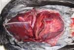
Image 89. Egg-bound African Grey (image courtesy Julie Burge; used with permission).
17.2.1 Late Hatch
Late hatch can be attributed to the old age of the parents, according to K. Joyner. “If ovulation ceases suddenly due to trauma or stress, then developing follicles may hemorrhage, resulting in regression of the developing follicle. Aging hens can exhibit permanent ovarian involution, which is believed to be a normal physiological process.” 32
17.2.2 Cystic Ovarian Disease
Cystic ovaries may occur alone or with other conditions, such as egg-yolk peritonitis and ovarian neoplasia. Other diseases which affect mature reproductive hens and senior hens include peritonitis, egg-yolk stroke and oviductal prolapse. “It may be asymptomatic or be accompanied by symptoms such as abdominal distention and dyspnea due to the pressure within the abdominal cavity. Removal of the fluid that has built up may relieve the discomfort. Leuprolide acetate has been used to stop production of ova.” 37 Diagnosis is by radiology or ultrasound.
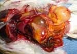
Image 90. Cystic ovary (image courtesy Julie Burge; used with permission).

Image 91. Prolapse due to egg-binding in a budgie (image courtesy Julie Burge; used with permission).
17.2.3 Cloacal and Uterine Prolapses
According to Bob Doneley, cloacal and uterine prolapse are caused by:
- Excessive straining
- Masturbatory behavior in male cockatoos
- Sexual overwork
- Oviductal disease, such as:
- Egg binding
- Salpingitis
- Intra-abdominal pressure
- Cloacal diseases such as cloacoliths (solid masses of debris) and cloacitis (inflammation or infection of the cloaca)
- Internal papillomas
- Internal papillomas
- Constipation or diarrhea 15
In geriatric birds, prolapses are generally found in the females and are related to loss of cloacal muscle tone due to chronic trauma from excessive breeding, difficulty in egg-laying, and retained and laying of soft-shelled eggs over the lifetime of the bird. 15
“The bird presents with a protrusion of the cloacal mucosa (with or without rectum or oviduct) through the lips of the vent. The prolapse is usually accompanied by straining and grunting while the bird is defecating, and she may pass blood around the vent or in the droppings.” 15 If the prolapse is not treated immediately, the tissue may be filled with fluid, and necrosis can result. To treat it, the practitioner will reduce the prolapse and place it back into the cloaca. If the cause is not found and dealt with, prolapses will probably recur. 15


Image 92. Prolapsed cloaca on an umbrella cockatoo (image courtesy Wilson and Lightfoot). 62
Image 93. Prolapsed uterus in a budgerigar (image courtesy Doneley)16

Image 94. Uterine prolapse, including partial prolapse of the vagina and cloaca in a 7-year-old cockatiel with a history of chronic egg-laying (image courtesy H. Bowles).7
18 Decline in Activity in the Senior Bird
18.1 Physical Decline Due to Malnutrition
Geriatric birds are more susceptible to certain health issues. Since malnutrition and nutritional disorders are still common in pet birds, and many owners continue to feed a seed-only diet, these birds will develop long-term health problems, including hypovitaminosis A, calcium deficiency, hepatic lipidosis, and secondary infection. Birds can live a very long time on these deficient diets without any outward signs of malnutrition, but over time it takes its toll. Even if the bird is changed to a better diet, sometimes it’s too late, and the bird has developed illnesses that cannot be helped. Also, even though the owner may offer better foods, the bird may not adapt to this new diet and only choose foods he is used to or enjoys. 65
18.2 Mental decline
18.2.1 Senility and Dementia
T. Lightfoot states that the effects of aging on birds’ cognitive abilities are not known at this time. She discusses studies that have shown that parts of the brain, the hippocampus in particular, are reduced in tissue volume in captive birds. This leads to memory loss. “Although no controlled studies of senility in birds have been reported, there are a few anecdotal reports from practitioners and owners of older birds which suggest that some aspects of mental decline may occur.” 37 The birds may not be able to locate certain places, such as his cage door and food dishes, even though they had good vision and had always found these easily in the past. Also noted were behavioral changes. “The birds began to interact differently with their long-term human and avian companions. These changes could be related to their mental state, but also could be caused by pain, lack of good muscle control, or bone health.” 37
19 The Geriatric Bird’s Physical Exam
A complete work-up and physical exam should be done annually, particularly if the older bird has ailments. He may even need to go in more frequently. He should not be anesthetized if at all possible, since older birds have a greater risk of complications. The practitioner should conduct a thorough physical exam at each visit and handle the bird very gently. 64
The annual office visit gives the owner and practitioner the opportunity to prevent disease by correcting husbandry issues. They are able to discuss any symptoms that might be early warning signs of disease. 23 The practitioner should include in his discussion:
- Changes in behavior that could a sign of pain, nervousness, fear or disorientation
- The extent of vocalization, play, and movement within the bird’s environment
- Interaction with objects, other birds, and people
- Quality-of-life concerns; these should be compared to previous years
- Changes in water consumption or urinary/fecal/urate output
- Changes in food preferences or eating habits
- Changes in preening habits, perching locations, and sleep cycles [10,23,37,64]
19.1 The Thorough Physical Exam Should Include:
- Taking and recording the weight of the bird
- Oral and fecal Gram’s stains
- Inspecting under the wings and around the vent
- Auscultating the air sacs, heart, and lungs
- Testing for muscle mass, exercise tolerance, and grip strength
- Range-of-motion testing for joints and limbs, walking gait, and flight, if possible
- Scrutinizing the feathers and skin for evidence of depigmentation (which may indicate a delayed molt), lack of preening (as evidenced by dirty, ratty, unzipped feathers), feather picking or chewing, or other abnormalities
- Examination of the eyes, ears, nares, oropharynx, choana, tongue and glottis
- Palpation of the crop and abdomen
- Visual examination of the uropygial gland (when present), and gentle massaging to express the sebum
- Examination of the cloacal mucosa
- Checking the feet for lesions and palpation of the feet, toes, and nails [10,23,37,60,64]
19.2 The need for additional testing
If the practitioner determines additional tests are necessary, they should include:
- Radiographs, to evaluate bone density and joint disease, arthritic changes, lesions, cardiovascular abnormalities, organ enlargement, and to screen for any internal masses
- Complete blood count (CBC) and plasma chemistries, including bile acids, cholesterol and HDL and LDL levels if indicated
- Ophthalmic examination
- Echocardiogram and ECG if warranted
- Skin tests, such as impression tests, skin scraping, or fine-needle aspirate of any growths
- Bacterial cultures and sensitivities 10,26,37,64
20 Summary of Diseases and Illnesses Frequently Seen in Geriatric Birds
- Atherosclerosis, mostly seen in African Greys and Blue-fronted Amazons, usually diagnosed on post-mortem.
- Strokes and heart attacks
- Skin diseases or conditions
- Fungal infections, such as aspergillosis, which may occur in older birds, especially those housed in poor conditions
- Chronic diseases of the liver, heart, or kidneys
- Neoplasia, most frequently seen with budgies
- Nutritional deficiencies, usually hypovitaminosis A. Dietary correction should not be undertaken until the bird has been examined, tested and treated for any illnesses. Stressing a bird by changing its diet can create an acute illness from sub-clinical disease.
- Swollen choana, and blunted or absent choanal papillae. Sheets of sloughed epithelial cells may be seen on a Gram’s stain
- Chronic sinusitis, often complicated by secondary bacterial or fungal infections
- Obesity, occurring when the weight exceeds the optimum by 15% or greater. Birds become more sedentary as they age, and the lower activity level may result in weight gain. The older bird has a slower metabolic rate, and may not require as many calories. Although hypothyroidism is not well-documented in psittacines, it may also play a role in weight gain in some older birds. [44,63,64]
- Weight loss. A loss of more than 10% of a bird’s body weight can mean illness. The bird should be seen immediately.
Conclusion
Most of the birds which were imported or hatched in the 1970’s and 1980’s have already passed away. However, a new group of healthier birds is moving into senior status at this time—birds hatched as 2nd, 3rd and 4th generation or more—captive-bred psittacines. Small birds whose lifespans a few years ago were only 10 years, and large birds whose lifespans were only 25-30 years, are now living to be 30 to 40 years and 80-or-more years, respectively.
Avian veterinarians are improving their skills and knowledge bases by attending on-going education conferences and seminars. Keeping up with all the new findings is challenging for busy clinicians, but they are devoted to their work, clients, and patients. An increase in clinical research into diseases has led to improved testing, diagnoses, medications, and treatments of these birds. Veterinarians are educating their clients about their geriatric birds’ special needs and potential illnesses. Improving client education in nutrition, husbandry, and preventative health care has become the goal of the practitioner, and as owners have become educated, they are more willing to procure quality veterinary care for their birds. This has led to healthier lifestyles and consequently, extended longevity rates in captive birds.
In the past, avian veterinarians have not needed to delve into geriatric medicine, but now that they are seeing more and more elderly birds in their practices, it is becoming necessary for them to study this new phase of life. As many as 30% of some practices are comprised of geriatric patients, so the need for more education in this field by the avian veterinarian and his clients has become a necessity. More and more owners are now dealing with the concerns surrounding old age in their birds—including quality-of-life issues. Although bird care is far improved from the past decades, the time has come for both avian specialist and bird owner to address the wellbeing of the geriatric psittacine. The annual check-up is the best time to do this, and it should be done before the bird reaches old age so the owner is prepared for changes in his bird’s health.
In Gratia
The author is deeply indebted to Drs. Robert Dahlhausen, Julie Burge, Peter Wilson, Zofia Evangeline Sangushko, Bart Huber, and the lay contributors for permission to use their images in this paper. Special thanks to Dr. Dahlhausen for reviewing and approving this paper.
1. Aydinonat D et al. Social Isolation Shortens Telomeres in African Grey Parrots (Psittacus erithacus erithacus). Abstract. PLoS One 2014 Apr 4:9(4):e93839. doi: 10.1371/journal.pone.0093839. eCollection 2014. – PubMed –
2. Austad S. Birds as Models of Aging in Biomedical Research. ILAR J 1997;38(3):137-141.
3. Austad SN. Candidate Bird Species for Use in Aging Research. In: Experimental Gerontology 2011, p. 36. From: ILAR Journal. Vol. 52, No. 1, 2010. – PubMed –
4. Bauck L. Mycoses. In: Avian Medicine: Principles and Application, Spix Pub., CD Rom. 2006 p. 1001.
5. Bennett R. Neurology. In: Avian Medicine: Principles and Application, Spix Pub., CD Rom. 2006, p. 739.
6. Bennett R and Harrison G. Soft Tissue Surgery. In: Avian Medicine: Principles and Application, Spix Pub., CD Rom. 2006, pp. 1099, 1100.
7. Bowles HL. Evaluating and Treating the Reproductive System. In: Clinical Avian Medicine Spix Pub., CD ROM 2006. pp. 525,529, 534.Available from www.ivis.org
8. Bowles HL. Surgical Resolution of Soft Tissue Disorders. In: Clinical Avian Medicine Spix Pub., CD ROM 2006. pp. 794, 827.Available from www.ivis.org
9. Brue RN. Nutrition. In: Avian Medicine: Principles and Application, Spix Pub., CD Rom. 2006, pp. 64, 72
10. Burkett G. Aging in Psittacines. In: The Birdie Boutique. www.birdieboutique.com/noname.html
11. Campbell T. Cytology. In: Avian Medicine: Principles and Application, Spix Pub., CD Rom. 2006 pp. 214, 218, 316
12. Clubb S and Karpinski L. Aging in Macaws. In: Proceedings of the Assoc Avian Vet 1992. pp. 83-86.
13. Dahlhausen RD. Implications of Mycoses in Clinical Disorders. In: Clinical Avian Medicine, Spix Pub., CD-ROM 2006 pp. 694-696.Available from www.ivis.org
14. Degernas LA. Trauma Medicine. In: Avian Medicine: Principles and Application, Spix Pub., CD Rom. 2006 pp. 425, 426
15. Doneley B. Avian Medicine and Surgery in Practice: Companion and Aviary Birds. Manson Pub. 2011, pp. 142-144, 169, 627. – Available from amazon.com
16. Doneley B, Harrison G and Lightfoot T. Maximizing Information from the Physical Exam. In: Clinical Avian Medicine, Spix Pub., CD-ROM 2006 pp. 169, 170, 190, 199, 201, 204, 205.Available from www.ivis.org
17. Echols MS. Evaluating and Treating the Kidneys. In: Clinical Avian Medicine Spix Pub., CD ROM 2006 p. 458.Available from www.ivis.org
18. Fitzgerald BC, Beaufrere H. Cardiology. In: Current Therapy in Avian Medicine and Surgery, 1st Ed., Brian l. Speer, (Ed.) Elsevier Pub. 2016 pp. 265-271, 288. – Available from amazon.com
19. Gerloch H. Bacteria. In: Avian Medicine: Principles and Application, Spix Pub., CD Rom 2006 p. 953.
20. Greenacre CB. Birds of a Certain Age–Overview of Geriatric Diseases. In: Proceedings of the Ass of Avian Veterinarians, 2014 pp. 150-152.
21. Greenacre CB. Necropsy Examination. In: Avian Medicine: Principles and Application, Spix Pub., CD Rom. 2006 p. 363.
22. Harrison G and McDonald D. Nutritional Considerations, Section II. In: Clinical Avian Medicine Spix Pub., CD ROM 2006 pp. 124, 130, 133, 134.Available from www.ivis.org
23. Harrison GJ and Ritchie BW. Making Distinctions in the Physical Exam. In: Avian Medicine: Principles and Application, Spix Pub., CD Rom 2006 p. 172.
24. Helmer P and Redig P. Surgical Resolution of Orthopedic Disorders. In: Clinical Avian Medicine, CD Rom, Spix Pub. 2006 p. 771.Available from www.ivis.org
25. Hillyer E. Gastroenterology (Image). In: Avian Medicine: Principles and Application, Spix Pub., CD Rom. 2006 p. 495.
26. Hochleithner M. Biochemistries. In: Avian Medicine: Principles and Application, Spix Pub., CD Rom. 2006 p. 25.
27. Hochleithner M, Hochleithner CH and Harrison LD. Evaluating and Treating the Liver. In: Clinical Avian Medicine, CD Rom, Spix Pub. 2006 pp. 440, 441, 442.Available from www.ivis.org
28. Holmes DJ, Fluckiger R and Austad SN. Comparative Biology of Aging in Birds: An Update. Exp Gerontol 2001 Apr;36(4-6):869-83. – PubMed –
29. Holmes DJ and Ottinger MA. Aging in birds. AGE 2005; 27: iii-v.
30. Hoppes SM. Geriatric Diseases of Pet Birds. In: Merck Veterinary Manual, 2015. SE Aiello, MA Moses, (Ed’s). Merck Sharp and Dohne Corp. Publ.Available online
31. Huber Bart. Personal communication, images, and text.
32. Joyner K. Theriogenology. In: Avian Medicine: Principles and Application, Spix Pub., CD Rom 2006 pp. 749, 771, 791.
33. Korbel R. Ophthalmology (Image). In: Avian Medicine: Principles and Application, Spix Pub., CD Rom 2006 p. 693.
34. Latimer KS. Oncology. In: Avian Medicine: Principles and Application, Spix Pub
35. Latimer K.S., Rakich P.M. Necropsy Examination. In: Avian Medicine: Principles and Application, Spix Pub., CD Rom 2006 pp. 316, 370
36. Latimer S.J. Internal Medicine. In: Avian Medicine: Principles and Application, Spix Pub., CD Rom. 2006, p. 662, 653, 720
37. Lightfoot Teresa L. Geriatric Psittacine Medicine. In: Veterinary Clinics of North America, Exotic Animal Practice, Geriatrics. Sharman Hoppes and Patricia Gray, (Ed’s.) Saunders/Elsevier, Inc. Publ. Vol. 13, No. 1. Jan. 2010 pp. 27-49, 32, 34, 38
38. Lightfoot T. Overview of Tumors: Section I. In: Clinical Avian Medicine, CD Rom, Spix Pub. 2006 p. 560, 561
39. Lumeij J.T. Endocrinology. In: Avian Medicine: Principles and Application, Spix Pub., CD Rom. 2006, pp.591, 592, 594
40. Lumeij . J.T. Gastroenterology. In: Avian Medicine: Principles and Application, Spix Pub., CD Rom. 2006 p. 495
41. Lumeji, J.T. Nephrology. In: Avian Medicine: Principles and Application, Spix Pub., CD Rom. 2006 p. 540
42. Lumeij J.T. Hepatology. In: Avian Medicine: Principles and Application, Spix Pub., CD Rom 2006 p. 529
43. Lumeij J.T., Ritchie BW. Cardiology. In: Avian Medicine: Principles and Application, Spix Pub., CD Rom. 2006 pp. 719-720
44. Macwhirter P. Malnutrition. In: Avian Medicine: Principles and Application, Spix Pub., CD Rom. 2006 p. 144
45. McMillan M.C. Imaging Techniques. In: Avian Medicine: Principles and Application, Spix Pub., CD Rom. 2006 pp. 316, 324
46. Miesle J. Resolution of Xanthomatosis without Surgical Intervention. On: Beauty of Birds
Website: www.beautyofbirds.com/xanthoma.html, and Facebook group: The Science of Avian Health in the Files, and on the IVIS website. 2013
47. Miesle J. Nutritional Requirements of Psittacines. On: Beauty of Birds website: www.beautyofbirds.com/NutritionalRequirementsofParrots.html and on Facebook group: The Science of Avian Health, in the files, and on the IVIS website.
48. Miesle J. Pododermatitis. On: Beauty of Birds website: www.beautyofbirds.com/pododermatitis.html and in The Science of Avian Health Facebook group in the files, and on the IVIS website.
49. Miesle J. The effect of tobacco smoke on avian species. In: The Science of Avian Health Facebook group in the files, and on the IVIS website.
50. Pees M., et al. Evaluating and Treating the Cardiovascular System. In: Clinical Avian Medicine, CD Rom, Spix Pub. 2006 p. 390, 391, 393
51. Pendl H., Tizard I. Immunology. In: Current Therapy in Avian Medicine and Surgery, 1st Ed. Brian l. Speer (Ed.) Elsevier Pub. 2016 p. 412
52. Petrak M.L., Minsky L. Metabolic and Miscellaneous Conditions. In: Diseases of Cage and Aviary Birds, 2nd Ed., M.L. Petrak, (Ed.) 1982. Lea and Febiger Pub. p. 639.
53. Quesenberry K., Hillyer E. Supportive Care and Emergency Treatment. In: Avian Medicine: Principles and Application, Spix Pub., CD Rom. 2006 p. 411
54. Reavill Drury R., Dorrestein Gerry M. Pathology of Aging Psittacines. In: Veterinary Clinics of North America, Exotic Animal Practice: Geriatrics:. Sharman Hoppes and Patricia Gray, (Ed’s). Saunders/Elsevier, Inc. Publ. Vol. 13, No. 1. Jan. 2010 pp. 135-150.
55. Samour J. Management of Raptors. In: Clinical Avian Medicine. CD Rom, Spix Pub. 2006 pp. 588, 939, 946
56. Sandmeier Peter, Coutteel Peter. Management of Canaries, Finches and Mynahs. In: Clinical Avian Medicine. CD Rom, Spix Publ. 2006 pp. 909, 910
57. Schmidt R., Lightfoot T. Integument. In: Clinical Avian Medicine, CD Rom, Spix Pub. 2006 pp. 403, 405
58. Styles D. Metabolic, Neoplastic, and Nutritional Diseases of Birds. Proc North American Veterinary Conf, 2005 pp. 1232-1233
59 VCA Hospital. Cataracts in Birds. (Image). http://www.vcahospitals.com/main/pet-health-information/article/animal-health/cataracts-in-birds/810
60. Williams David. Ophthalmology. In: Avian Medicine: Principles and Application, Spix Pub., CD Rom. 2006, p. 693
61. Wilson L, Linden P, Lightfoot T. Early Psittacine Behavior and Development. In: Clinical Avian Medicine, CD Rom, Spix Pub. 2006 p. 60
62. Wilson L, Lightfoot T. Concepts in Behavior, Section III: Pubescent and Adult Psittacine Behavior. In Clinical Avian Medicine, CD Rom, Spix Publ. 2006 p. 76, 83
63. Wissman M. Fatty Liver Disease. Exotic Pet Vet. http://www.exoticpetvet.net/dvms/fattyliver.html
64. Wissman M. Growing Old Gracefully. Exotic Pet Vet. http://www.exoticpetvet.net/avian/geriatric.html
65. Wissman Margaret, Atherosclerosis. ExoticPetVet.net. 2006 http://www.exoticpetvet.net/avian/geriatric.html
Appendix A. Aging in Macaws – A Study by Susan Clubb
In 1992, Susan Clubb submitted a paper to the Association of Avian Veterinarians for publication and presentation at the annual conference proceedings. This paper, “Aging in Macaws,” documented the review of health records of a group of macaws whose ages were known in order to provide information about the aging process and longevity of macaws in captivity and in the wild.
She states that macaws are known to be “hardy and long-lived… with few natural predators,” 12 and she estimates their natural lifespans are at least 60 years. At the time, however, there had been very little documentation of their life spans or aging processes in academic literature. Her work was done at Parrot Jungle and Gardens in Miami, Florida, which had been in existence since 1936.
The 57 birds used in this study were at least 25 years old and were examined yearly for a period of three years. Twenty-six were captive-bred and ranged from 25 to 52 years of age. Thirty-one were wild-caught birds which had lived at the facility for at least 20 years. The oldest bird there at the time, a 57-year-old military macaw, had been recently euthanized due to frequent and severe seizures. It had been blind and had suffered from a chronic neurological disorder for many years.
As of the writing of Clubb’s paper in 1992, the first macaw that had hatched at Parrot Jungle, and was still in the collection, was 52 years old. Seven other captive-bred birds in the collection were between 41 and 49 years old. “All had various stages of cataracts and degenerative diseases related to senescence, including arthritis, loss of skin tone and elasticity, muscle wasting, and degenerative neurological disease. As with most mammalian species, aging macaws show physical signs of aging around the time that their reproductive potential has passed. Macaws are known to be capable of reproducing from four to thirty-five years of age. The most productive years were the late teens and early twenties.”
After 40, the onset of all degenerative changes began to occur. For the birds in this collection, lack of exercise led to weight loss and muscle wasting since these birds were sedentary. “Birds which had always been free-fliers reverted to walking or resting in a secure location most of the day.” Clubb states that their posture had not significantly changed; most remained upright when perching unless they had had skeletal injuries in the past. Arthritis, with its joint stiffness and limited range-of-motion, was evident in birds over 40 years of age. “Some birds over 40 developed twisting deformities of the flight feathers of the wings, similar to the Angel Wing disorder.”
Since macaws have no feathers on their faces, dermatological changes were obvious in birds over 40. “The skin on the face developed pigmented spots, polyps, wart-like blemishes, cysts, and wrinkling. The skin on the face and feet became thin, and the facial feathers at the edge of the skin began to thin out; in addition, the skin on the feet began to develop spotty depigmentation. Feathers in general began to lose condition and luster at this point as well.”
Most macaws developed degenerative eye disorders, primarily cataracts, after age 35. “The external ophthalmic changes included lid laxity. The lids lost tone and became thickened and wrinkled. The corneas remained relatively clear unless there had been some interocular trauma in the past. Most birds in this collection were blind in at least one eye after age 45.” These older macaws in this collection died as a result of tumors, renal failure and other degenerative diseases. “Many had died long before their natural lifespans has been reached due to malnutrition and husbandry-related disease. Once a bird develops cataracts and is unable to see well, his survivability in the wild is limited since cataracts limit his ability to forage.” Most macaws over 35 years of age developed cataracts, just as they develop in other species of birds. “Cataracts may develop suddenly, and this can lead to uveitis and blindness. In macaws, age-related cataracts are seen in the center of the lens. Surgery to remove them is possible and sometimes performed.”
What is interesting is that these birds were tracked in the late 1980’s to early 1990’s, and at that time, the macaw’s maximum lifespan was only in the 40’s and 50’s. Since then, only a little over 20 years later, due to improved diet and husbandry practices, the maximum lifespan has extended to 50-90 years of age in some species.
Appendix B. Carly, an aged macaw.
(images approved by Pete DiSalvo; used with permission)
The following set of pictures is of a 75-year-old scarlet macaw named Carly. She was passed down in the family, and recently the last person who had her passed away. She was adopten by Pete DiSalvo of Sugarcreek Bird Farm in Bellbrook, Ohio in late 2016. She is a most interesting study in the effects of aging on macaws. Decades of neglect, poor nutrition, and poor perching have taken its toll on this bird.


Image 1. The plantar surface of the feet is surprisingly free of pododermatitis. There are some areas that are a little shiny and rough, but hopefully good nutrition and better places to stand will help. The bird has severe arthritis and has been standing instead of perching for many years. She stands very low to the surface of her platform because of her arthritis. Mr. DiSalvo has placed the bird in a large cage with a platform instead of a perch. He will put padding on the platform to give her a softer surface.
Image 2. Note the poor condition of the feathers. As feather follicles age, they either stop producing feathers completely or produce feathers which have poor pigmentation and/or poor and missing barbs. Also note the condition of the nails. Some of the digits are useless and cannot grip at all. Most are curved and twisted away from the toe, and the toes are misshapen.
Image 3. The plumage doesn’t appear to be too bad on the back, but up close, missing, discolored, and twisted feathers can be seen. Her stance is remarkably erect, in spite of her difficulty with arthritis. She usually leans against something, though, for support.


Image 4. Carly’s face is remarkably free of dermatolotical disfigurement. She has no lumps, warts, or other markings typical of such an old bird. Note that her maxilla is now in good condition after having been dremmeled down by Dr. Dahlhausen. It was reaching almost to the neck. The mandible is shaped like a bowl because of the pressure on it from the maxilla. She was developing a scissors beak. Dr. Dahlhausen did an extraordinary job getting the beak in this condition considering what Carly had been going through just to eat. There is significant improvement in her bite now.
Image 5. The breast shows the decrease in feather color and condition due to age, poor nutrition, and neglect. Again, note the feet. The toes are nearly useless from inability to grip, and the nails curved in all directions.
Image 6. Pete DiSalvo, the kind, caring man who took Carly in. His generous heart has been repaid tenfold because of the love he receives from this sweet bird. He’d do it all again.