Aspergillosis is an infectious, non-contagious fungal disease caused by species in the ubiquitous opportunistic saprophytic genus Aspergillus, in particular Aspergillus fumigatus.

by Dr. Tara Nath Gaire
Department of Veterinary Medicine and Public Health
Institute of Agriculture and Animal Science (IAAS), Rampur, Chitwan, Nepal.
Index of Bird Diseases … Symptoms and Potential Causes … Article: The Silent Parrot Killer / Aspergillosis in Pet Birds
Synonyms: Brooder pneumonia, Mycotic pneumonia, Pneumomycosis
Abstract
Aspergillosis is an infectious, non-contagious fungal disease caused by species in the ubiquitous opportunistic saprophytic genus Aspergillus, in particular Aspergillus fumigatus. This mycosis was described many years ago, but continues to be a major cause of mortality in captive birds and, less frequently, in free-living birds. Although aspergillosis is predominantly a disease of the respiratory tract, all organs can be involved, leading to a variety of manifestations ranging from acute to chronic infections. It is believed that impaired immunity and the inhalation of a considerable amount of spores are important causative factors. The pathogenesis, early diagnostic methods and antifungal treatment schedules need to be further studied in order to control this disease. The aim of the present review paper is to present the current knowledge on aspergillosis in domestic poultry. The review covers etiology, epidemiology, pathogenesis, clinical signs and lesions, diagnosis, treatment control and prevention of Aspergillosis.
Key words: Aspergillus, Aspergillus fumigates, Brooder pneumonia
FREE Training to Stop Your Bird's Biting
Get FREE access to Beak School – the only system designed to train your bird. Stop behavioral problems like biting, screaming, feather plucking.
Get the Free TrainingIntroduction
The commercial poultry industry in Nepal is growing tremendously. The contribution of poultry to the national GDP is nearly 3% of total GDP and 8% of the agriculture GDP of the country (Dhakal, 2000). Poultry production in Nepal started since last four decades and now it has been already recognized as one of the most dynamic sector and has taken the position of an industry. A dramatic growth rate in chicken population has been observed since then. The introduction of highly productive breed of poultry, balanced nutrition, modern housing and management practices, disease control measures, adaptation of modern technology and improved marketing system has contributed for the development of poultry industry to develop as a major sector of agribusiness. Together with the increase in commercialization of the industry, more and more diseases are becoming prevalent. Poultry farming has been suffering from many diseases mainly due to lack of biosecurity in the farms, technical knowledge among farmers, faulty shed construction, substandard health and management practices, poor quality of chicks and feeds and environmental and climatic constraints.
Aspergillosis is a disease, usually of the respiratory system, of chickens, turkeys, and less frequently ducklings, pigeons, canaries, geese, and many other wild and pet birds. In chickens and turkeys, the disease may be endemic on some farms; in wild birds, it appears to be sporadic, frequently affecting only an individual bird. It is usually seen in birds 7-40 days old. Chicks and poults may become infected during hatching as a result of inhaling large numbers of spores in heavily contaminated hatching machines or from contaminated litter. In older birds, infection is caused primarily by inhalation of spore-laden dust from contaminated litter or feed or dusty range areas.
In spontaneous outbreaks, mortality ranged between 4.5% and 90%, whereas age of diseased birds varied from 3 days to 20 weeks. Beside direct losses related to mortality, feed conversion and growth rate in recovering birds remain poor. Indeed, airsacculitis is a major reason for carcass condemnation at slaughter inspection.
Etiology
Aspergillosis is mostly caused by Aspergillus fumigatus but Aspergillus flavus, Aspergillus niger, Aspergillus glaucus,Aspergillus nidulans, and other Aspergillus species or mixed infections can play a role in the disease (Barton et al.,1992; Perelman and Kuttin, 1992; Joseph, 2000). The reason why A. fumigatus is the predominant species of airborne fungal infections might be that the spores are much smaller than the spores of other Aspergillus species (Richard and Thurston, 1983).
Growth
The fungus grows quite readily on ordinary laboratory culture media at room temperature, at 37oC, and higher. Czapek’s solution agar or Sabouraud’s agar may be used. The colonies are green to bluish green at first and darken with age so as to appear almost black. The colonies vary from velvety to floccose. The conidiophores are short, up to 300µ long by 2-8µ in diameter, the vesicles are apical flask-shaped up to 20-30µ in diameter, the sterigmata are 6-8 by 2-3µ in diameter, in chains forming solid columns up to 400 by 50µ (Thom and Church, 1926).
Toxins
Leber, according to Lucet (1897) and Van Heelsbergen (1929), succeeded in isolating toxins from cultures of Aspergillus fumigatus. Ceni and Besta (1902) were able to extract toxic materials from spores. A toxin reported by Bodin and Gautier (1906) was similar to bacterial toxins and produced clonicotonic convulsions, paralytic symptoms, and finally death. A toxin obtained by Henrici (1939) was toxic for rabbits, guinea pigs, mice, and chickens. This toxin was hemotoxic, neurotoxic and histotoxic. Rabbits and dogs are very susceptible to Aspergillus toxin. Pigeons, however, which are very susceptible to spontaneous infection, are very resistant to injected toxin.Japanese workers Asakura et al. (1962) studied an allergic skin reaction against an alcoholic precipitate from a mycelial extract of A. fumigatus. Penguins showed severe and prolonged skin sensitivity, whereas pigeons and ducks were quite resistant. Zoo penguins frequently die from A. fumigatus infections.
Forgacs and Carll (1955) selected strains of Aspergillus, Penicillium, and Alternaria isolated from samples of feed associated with outbreaks of a hemorrhagic disease in poultry, and grew the cultures on grain which was fed to day-old chicks. Subsequently the chicks developed hemorrhages of the muscles, lungs, heart, gastrointestinal tract, and liver. Twenty chicks 6 weeks of age were given feed on which the species of Alternaria had been cultured alone; they died and the necropsy revealed hemorrhages throughout the carcass.
Epidemiology
Aspergillosis is encountered in poultry in two main forms. Acute outbreaks in which there are high morbidity and high mortality may occur particularly in young birds. In adults especially, an occasional bird in a flock or aviary may become affected while the other birds remain healthy. The numerous reports in the literature suggest that nearly all species of birds may be affected.An increased concentration of spores in the environment may predispose a bird to aspergillosis. A warm environment, humidity, poor ventilation (Phalen, 2000; Tell, 2005), poor sanitation (Oglesbee, 1997) and the long-term storage of feed (Khosravi et al., 2008) may increase the amount of spores in the air. Factors impairing the bird’s immunity can also predispose to mycosis. Examples of this include the administration of tetracyclines (Oglesbee, 1997) or long-term steroids (Verstappen and Dorrestein, 2005), vaccination (Barton et al., 1992), metabolic bone disease (Vanderheyden, 1993), an inadequate diet (Bauck et al., 1992; Dorrestein, 1992) resulting in hypovitaminosis A (McMillan and Petrak, 1989; De Herdt, 1996), overcrowding (McMillan and Petrak, 1989), shipping (Tsai et al., 1992), quarantine or capture of wild birds (Abrams et al., 2001), starvation, thermal discomfort, migration (Young et al.,1998), inbreeding (Low et al., 2005), circovirus infection (Soike et al., 1999) and lymphoproliferative disorders (Kelly et al., 2004), toxicosis (Young et al., 1998; Carrasco et al., 2001; Jung et al., 2009), traumatic injuries (Xavier, 2008) and reproductive activity (Jones and Orosz, 2000).
Active fungal proliferation and sporulation of A. fumigatus on organic material produce large amounts of airborne small-sized conidia that are easily dispersed in air, then potentially inhaled and deposited deep in the respiratory tract. Susceptible hosts will develop polymorphic clinical forms in relation to either localized or disseminated lesions. Acute aspergillosis generally occurs in young birds resulting in high morbidity and mortality. The chronic form is sporadic. It causes lesser mortality and generally affects older birds, especially breeders in poultry, presenting a compromised immune system due to poor husbandry conditions.
Transmission
A case of egg-borne aspergillosis was reported by Eggert and Barnhart (1953). They suggested that the fungus had penetrated through the eggshell during incubation and the recently hatched chicks were infected. Another case which was shown to be hatchery-borne was reported by Clark et al. (1954). From 21 ranches where 210,000 chicks were involved, there was mortality of 1-10%. The infection could not be traced to the hatching eggs but was readily found in the incubators, hatchers, incubator rooms, and intake ducts. Symptoms and lesions were noted in some day-old chicks, but generally classical lesions were observed in chicks 5 days of age.
O’Meara and Chute (1959) produced aspergillosis experimentally in hatching chicks. Chicks in the process of hatching and up to 2 days of age were easily infected with A. fumigatus spores by contaminating the forced draft incubator with wheat which had been seeded with A. fumigatus. Chicks older than 3 days of age were resistant to infection.Chute and O’Meara (1961) reported some unusual cases of avian mycosis. Two cases of fungous tracheitis were observed, and in each case the mycelial growth extended through the cartilage
Pathogenesis
Inhalation is considered the main infection route for A. fumigatus in birds (Oglesbee, 1997), and because A. fumigates spores are too small to be trapped completely in the nasal cavity or trachea, some are able to reach the lungs and air sacs (Fedde, 1998). The air sacs are usually the primary infection sites, since inhaled air reaches the posterior thoracic and abdominal air sacs prior to contacting epithelial surfaces in the lungs (Nardoni et al., 2006). In the lung parenchyma, spores get embedded in the atria and parts of the infundibula in the parabronchus and are engulfed by (surface) phagocytic epithelial cells (Maina, 2002). When there are too many spores or the bird has an impaired immune response, the innate defence mechanisms do not succeed in eliminating infection at the site of the air capillaries. This may lead to the development of loosely attached plaques, which may or may not become overgrown by connective tissue of the host. These plaques or necrotic debris in the respiratory tract can obstruct the trachea or bronchi and/or fill up the air sacs (Oglesbee, 1997). Occasionally, sporulation occurs in the lungs and air sacs (Nardoni et al., 2006; Cacciuttolo et al., 2009). Hyphae containing fruiting bodies can fill the lumen and may penetrate the air sacs, causing serositis and superficial necrosis in the adjacent organs (Tsai et al., 1992). Besides direct extension of the infection through the air sac wall, disseminated mycosis also occurs by haematogenous spread. Hyphae, which are known to be tissue and angio-invasive (Dahlhausen et al., 2004), as well as host cells play a role in this spreading mechanism. Macrophages in the respiratory tract ingest spores and find their way through the interstitium into the blood and lymphatic stream and thus to other organs (Richard and Thurston, 1983).
The relevance of virulence factors in avian aspergillosis is not well known, as research on this is minimal (Peden and Rhoades, 1992; Richard et al., 1994, 1996). Ongoing studies suggest that A. fumigatus conidia may be able to resist killing by the avian respiratory macrophage (Van Waeyenberghe et al., 2009).
Two types of tissue reaction are recognized: the granulomatous or deep nodular form, and the infiltrative or superficial diffuse form. In the first type, neither exudative inflammation nor vascular lesions in the neighbouring tissues are seen. This type of encapsulated reaction develops both in non-aerated and aerated organs (Nardoni et al., 2006; Femenia et al., 2007; Cacciuttolo et al., 2009). In the non-ridged, non-encapsulated infiltrative type, the fungus frequently invades blood vessels. In aerated organs such as the lungs and the air sacs, the fungus may form aggregates of radiating hyphae containing large numbers of conidiophores and conidia in the absence of a structured granuloma formation (Nardoni et al., 2006; Cacciuttolo et al., 2009). A mixed type composed of both tissue reactions in the same tissue section is also possible (Tsai et al., 1992; Atasever and Gümüssoy, 2004).
The popular assumption that the paucity of free avian respiratory macrophages results in an inadequate or incompetent avian respiratory immune system and is therefore responsible for the high susceptibility of birds to respiratory pathogens (Toth and Siegel, 1986) lacks scientific foundation. Instead, the susceptibility of birds to Aspergillus spp. may be attributed to differences in anatomical, physiological and respiratory immune system characteristics compared with mammals (Toth, 2000; Maina, 2002; Tell, 2005).
Clinical Signs, Gross Lesions, Histopathology and Diagnosis
Clinical Signs
Clinical manifestations depend on the infective dose, the spore distribution, pre-existing diseases, and the immune response of the host (Dahlhausen et al., 2004). Avian aspergillosis is often classified as acute or chronic. Acute aspergillosis is thought to be the result of inhaling an overwhelming number of spores, while chronic aspergillosis is generally associated with immune suppression (Vanderheyden, 1993). Acute aspergillosis may include a variety of nonspecific clinical signs: anorexia, lethargy, ruffled feathers, respiratory signs, polydipsia, polyuria, stunting, or sudden death. In chicks, contaminated in ovo or during hatching, the disease, commonly known as brooder pneumonia, is highly fatal in the first ten days of life and results in a major respiratory distress. Two outbreaks of omphalitis where the primary cause was A. fumigatus have been investigated in young turkeys.In poultry farms, mortality rate may rise slightly or increases suddenly, peaks during a few days, and then returns to initial state Respiratory signs include dyspnoea, gasping, hyperpnoea with panting, nonproductive coughing, wheezing, cyanosis , and sometimes nasal discharge .In the chronic form, dyspnoea, depression, dehydration, and emaciation are described. Nervous system involvement causes ataxia, tremor, opisthotonos, lateral recumbency, torticollis, seizures, convulsions, lameness, and hind limb paresis .Occurrence of nervous and ophthalmic troubles one week after an acute episode of aspergillosis has been reported in a turkey flock. Cloudiness of the eye with severe conjunctivitis and turbid discharge were associated with paralysis in broiler breeders.
Although aspergillosis is predominantly a disease of the respiratory tract, any organ can be infected. Nasal aspergillosis causes exudative rhinitis (Tsai et al., 1992), possibly accompanied by malformation of the nostrils, beak and cere (Bauck et al., 1992). Mycotic keratitis can cause blepharospasm, photophobia, periorbital swelling, turbid discharge, swollen and adhered eyelids, cloudy cornea and cheesy yellow exudates within the conjunctival sac (Beckman et al., 1994; Hoppes et al., 2000). Neurological signs can be caused by Aspergillus spp. (Jensen et al., 1997). Epidermal cysts associated with A. fumigatus have been described in the comb of a silky bantam chicken (Suedmeyer et al., 2002). Aspergillus blepharitis and dermatitis involving the eyelids and the head have been described in a peregrine falcon–gyrfalcon hybrid (Falco peregrinus x Falco rusticolus) (Abrams et al., 2001). Right ventricular dilatation (cor pulmonale) due to pulmonary hypertension, with or without ascites, and congestion of the lungs caused by ventricular failure occasionally occurs (Julian and Goryo, 1990; Höfle et al., 2001). Birds suffering from aspergillosis do not always show respiratory problems.
Gross Lesions
The severity and the degree of development of the disease determine both morphology and extension of macroscopic lesions. Extensive involvement of the respiratory tract can occur before clinical signs are apparent. Typically, lesions consist of white to yellowish granulomas ranging from miliary (mm in diameter) to large roughly spherical granulomatous nodules (>2![]() cm) involving serosae and parenchyma of one or multiple organs. Single or multiple necrotic areas are visible on cut surfaces. The primary location of lesions is the air sacs and lungs although oesophagus, proventriculus, gizzard, small intestine, liver, kidney, spleen, skin, trachea, peritoneum, brain, eye, muscle, or heart may be involved. Lung parenchyma is either consolidated or has focal granulomas of varied size (Figure 1). When coalescing in air sacs, these masses form cheesy caseous plaques covering the thickened membranes and even obstructing the entire lumina where fungal sporulation may occur as evidenced by a grey-greenish velvet.
cm) involving serosae and parenchyma of one or multiple organs. Single or multiple necrotic areas are visible on cut surfaces. The primary location of lesions is the air sacs and lungs although oesophagus, proventriculus, gizzard, small intestine, liver, kidney, spleen, skin, trachea, peritoneum, brain, eye, muscle, or heart may be involved. Lung parenchyma is either consolidated or has focal granulomas of varied size (Figure 1). When coalescing in air sacs, these masses form cheesy caseous plaques covering the thickened membranes and even obstructing the entire lumina where fungal sporulation may occur as evidenced by a grey-greenish velvet.
 |
Figure 1: Numerous nodules in the lung of poultry with acute aspergillosis |
Nonulcerative or mildly ulcerative keratitis was reported in a turkey flock . Broiler pullets presented periorbital and eyelid swelling with cheesy yellow exudates in the conjunctival sac . Circumscribed white to greyish areas were observed in the cerebellum of broiler breeders and turkeys . Ribs of ostriches , sternum of broiler breeders , yolk sacs of poults and hip joints of turkeys constituted unusual locations of aspergillosis lesions.
The lesions depend considerably on the site of infection. Either localisation or generalisation may be observed. Individual lesions may be observed, for example, in the syrinx or in a single air sac. The lungs are most frequently involved. Pulmonary lesions vary from miliary nodules up to larger nodules.In some cases there may be localised hepatization, and in others grossly visible mycelial masses may be present in the air passages and bronchi. There may be generalised involvement of the air sacs. Occasionally a circular disc-shaped necrotic mass with a concave surface, loosely attached to which there is a circular more or less flat or convex plaque, may be observed. Various manifestations of the disease have been described. Lange (1914) recorded nodules in the lungs and the thoracic and abdominal cavities of chickens, ducks, geese, and pigeons. These nodules varied from pinhead or millet seed size up to the size of a pea. They were yellow in colour, of an elastically soft cartilaginous consistency, and homogeneously caseous. Individual nodules were noted on the intestinal serosa and in the parenchyma of the liver in a goose.
The nodules in the lungs of a turkey observed by Schlegel (1915) were pinhead to lentil size and were surrounded by an infiltrated or hemorrhagic corona with considerable hepatization. There were also grayish yellow, fibrin purulent disc- or plate-shaped masses of exudates 2-5mm thick on the pleura. Inflammation and detritus masses were present in the bronchi. The anterior thoracic, maxillary, and cervical air sacs contained yellow caseous flat discs and masses consisting of inflammatory exudates and mycelia. The left lower thoracic, upper posterior thoracic, and left abdominal air sacs were greatly distended. The walls of these air sacs were thickened and covered with a fur like growth of mold. Adjacent to the air sacs there were lentil-sized, knob-shaped, concentrically layered, turbid yellow, solid nodules composed of fibrin and mycelia. There were about 200cc of reddish turbid fluid in the abdominal cavity.
There were no circumscribed yellowish foci in the outbreak in chicks reported by Savage and Isa (1933). There was a diffuse grayish yellow infiltration in the lungs, with about one-third of each lung involved. Mortality was 90% in this outbreak.In pneumomycosis in a flamingo described by Mohler and Buckley (1904), the lungs were filled with nodules, and the mucosa of the bronchi was covered with membranous masses that consisted primarily of the fungous mycelium.
Archibald (1913) found gray round colonies of the fungus in the bronchioles in an ostrich, whereas in a case described by Jowett (1913) the lungs were covered with miliary foci. Lahaye (1928) states that Aspergillus glaucus may be the cause of a disease of the skin in pigeons, particularly in young birds. Any part of the body may e affected with yellow scaly spots. The feathers in the affected areas are dry and easily broken.
Durant and Tucker (1935) observed yellowish white nodules up to 5 x 8mm in the lungs of wild turkey poults being reared in captivity. The hyphae of the fungus also penetrated the tissue of the lung, and there was involvement of the adjacent air sacs.In canaries observed by De Jong (1912) there were small whitish yellow, crusty coatings on the tongue, palate, and aditus laryngis and in the trachea and syrinx. Caseous foci in the lungs and caseous coatings on the pleura and peritoneum were also observed.
Histopathology
The histological picture as described by Nieberle (1923) consists of focal pneumonia, multiple necrosis, and nodular formations which resemble tubercles. The diffuse pneumonic foci are indicative of fibrinous or catarrhal pneumonia. The alveoli, bronchioles, and bronchi are filled with mucus, stained fibrin, nuclear fragments, detritus, leucocytic and inflammatory cells, and mycelia. The mycelium penetrates the walls, and the surrounding pulmonary parenchyma shows an exudative cellular inflammation or necrosis. The tubercle-like nodules show in the center a radiating turf of hyphae surrounded by a reactive inflammatory wall which resembles granulation tissue. Foreign body giant cells are frequently observed. The fruiting organs (conidiophores, sterigmata, conidia) occur more frequently in the air sacs.
In turkey cases of ophthalmitis described by Moore (1953), the primary involvement was in the viterous humor and the adjoining tissues. In one turkey he observed the presence of mycelia in the crystalline lens. Haematoxylin-eosin stain is often augmented with periodic acid-Schiff, Grocott, and Gomori’s methenamine silver stains in order to display fungal elements in embedded tissue sections. The fluorescent optical brightener blankophor proves to be a valuable tool for demonstrating Aspergillus sp. hyphae .Based on histopathological difference a deep nodular form and a superficial diffuse form of aspergillosis. A well-organised granulomatous reaction develops in nonaerated parenchyma whereas a superficial diffuse form, containing fungal elements and a nonencapsulated pyogranulomatous reaction, predominates in serosae and lungs .Organised granulomas are clearly encapsulated by an outer thick fibrous layer whereas pyogranulomas lack clear borders. The first feature corresponding to late granuloma may also be an important sign of the chronic form of aspergillosis, especially in adults Pyogranulomas organization presents a centre with variable amounts of septate, dichotomously branching hyphae surrounded by a palisade of radially arranged foreign body giant cells, macrophages, heterophils, and lymphocytes. Phagocytized fungal elements are regularly observed in the eosinophilic cytoplasm of multinucleated cells. Lymphocytes may infiltrate the margins of the granuloma Progressive inflammation in lungs resulting in small granuloma coalescence induces more extensive lesions and may lead to parabronchial obliteration with necrotic eosinophilic material containing erythrocytes, degenerated heterophils, and exfoliated epithelial cells . Numerous conidiophores and free spores appear in granulomas that open to the air spaces of the respiratory tract. Typical granulomatous reactions associated or not with fungal elements have been observed in brain conjunctiva, liver, spleen, gizzard, small intestine, hip joints, and trachea.
Diagnosis
The signs of aspergillosis are non-specific, making diagnosis difficult (Dahlhausen et al., 2004). Moreover, no single test provides certainty. Diagnosis usually relies upon an accumulation of evidence from the history, clinical presentation, haematology and biochemistry, serology, radiographic changes, endoscopy and culture of the fungus (Jones and Orosz, 2000).The history of the bird can reveal a stressful event and/or some underlying environmental factors and/or an immunosuppressive condition or treatment (Jenkins, 1991). It may also reveal chronic debilitation, voice change or exercise intolerance (Oglesbee, 1997).The clinical signs depend on which form of aspergillosis the bird develops and which organs are involved (Jones and Orosz, 2000). Hence, aspergillosis should be included in the differential diagnosis of respiratory tract and systemic diseases (Jenkins, 1991; Jones and Orosz, 2000).
Results of haematology and plasma biochemistry can be considered indicative rather than diagnostic (Jones and Orosz, 2000). Leukocytosis of 20,000 to more than 100,000 white blood cells per microlitre (Jenkins, 1991; Oglesbee, 1997), heterophilia with a left shift (degenerative shift), monocytosis and lymphopenia have been described in aspergillosis cases (Forbes, 1992). In addition, non-regenerative anaemia, increased total protein and globulin fraction can be observed (Vanderheyden, 1993; Reidarson and McBain, 1995; Jones and Orosz, 2000). Acute infections often present an increase in ?-globulins, while chronic infections show an increase in ?-globulin and/or ?-globulin fractions . However, immunosuppressed birds may have hypoproteinaemia (Ivey, 2000; Cray et al., 2009a) and white blood cells may be in the normal range (Flammer and Orosz, 2008). Overall, changes in protein electrophoresis are non-specific, but can be useful to estimate disease progression and the response to therapy (Ivey, 2000; Cray et al.,2009a).
Serological tests have been developed to confirm an early and more definite diagnosis of aspergillosis (Peden and Rhoades, 1992). However, in the acute stage, antibody production trails behind antigen exposure by 10 to 14 days (Brown and Redig, 1994); and if the bird is immunosuppressed, the low antibody production results in false negative results (Redig, 1994). In these cases, detection of circulating Aspergillus antigen in the serum may be more helpful (Cray et al., 2006). In chronic cases in which antigen levels may be low, detection of antibodies may be useful (Jones and Orosz, 2000). A number of serological test methods have been applied in birds, including counter-immunoelectrophoresis, agar gel immunodiffusion and enzyme-linked immunosorbent assays—but, in general, negative serological tests do not rule out aspergillosis, and positive tests are only considered diagnostic when backed up by other evidence (Peden and Rhoades, 1992; Brown and Redig, 1994; Redig, 1994; Redig et al., 1997; Le Loch et al.,2005; Arca-Ruibal et al., 2006; Cray et al., 2006, 2009a,b).
Although radiographs may not be helpful, lateral and ventrodorsal views can be taken in a bird suspected of having aspergillosis. For those that are unlikely to survive anaesthesia, standing or perching lateral views as well as dorsoventral views are helpful (Jones and Orosz, 2000). The radiographic changes of pneumonia and consolidating airsacculitis are non-specific (McMillan and Petrak, 1989). Consistent with radiography, computed tomography scans reveal the extent of lesions—but the diagnosis of aspergillosis still requires identification by biopsy, smear or culture (Phalen, 2000).
Endoscopy is invasive and requires anaesthesia, but it allows the extent of the lesions to be seen as well as the progress of infection during treatment (Redig, 1994; Jones and Orosz, 2000). This technique enables evaluation of the entire respiratory tract. Tracheal endoscopy is useful for showing a single lesion, such as a thick white discharge or plaque occluding the trachea or syrinx (Jenkins, 1991; Marks et al., 1994; Redig, 1994). The lower respiratory tract is best evaluated by laparoscopy (Jones and Orosz, 2000). Endoscopy of the abdominal air sac can show a diffuse cloudiness or white or yellow plaques. In the event of sporulation, plaques are covered with green–gray pigmented mould. Samples for culture and cytology should be taken directly with biopsy forceps or via air sac lavage (Jenkins, 1991; Taylor, 1993; Oglesbee, 1997).On necropsy, the yellow, green or white granulomatous foci can be noted in chronic aspergillosis patients (Jenkins, 1991; Vanderheyden, 1993). Acute aspergillosis causes numerous miliary granulomatous foci (McMillan and Petrak,1989; Jenkins, 1991).
Definitive diagnosis requires demonstration of the presence of the organism by cytology or histopathology and its identification by culture (Dahlhausen et al., 2004). It is important to mention that isolation of the fungus alone does not confirm the infection because Aspergillus organisms are ubiquitous and can be contaminants (Jensen et al., 1997; Flammer and Orosz, 2008). However, an abundant culture from any organ should be regarded as diagnostic. On the contrary, a negative culture does not rule out aspergillosis (Redig, 1994; Jensen et al., 1997).
Histopathological lesions can be suggestive, but because in vivo hyphae of hyaline filamentous fungi are very similar and their in situ manifestations are not pathognomonic, this technique does not allow fungal species identification (Kaufman et al., 1997; Cray et al., 2009a). Thus the aetiological diagnosis should ideally be confirmed by immunohistochemistry, although few reports on immunohistochemical techniques using monoclonal or polyclonal antibodies in birds with aspergillosis are available (Carrasco et al., 1993; Jensen et al., 1997; Beytut et al., 2004, Beytut, 2007).A few reports of different polymerase chain reaction assays (including real-time polymerase chain reactions) tested on heparinized whole blood, tracheal washings, air sac fluids, respiratory tract granulomas, or (biopsy) tissue samples from birds support the value of this assay in diagnosing avian aspergillosis (Dahlhausen et al., 2004; Cray et al., 2009a). However, further research is necessary before such assays can be included in the work-up of the avian practitioner.
Treatments and Acquired Immunity
Treatments
Treating avian aspergillosis is a challenge due to a number of factors. These include the limited knowledge on the pharmacokinetics of antifungal agents in different bird species, the granulomatous inflammation that makes it difficult for the drug to reach the target fungus, the presence of concurrent disease and/or immunosuppression, and the late stage at which birds are usually presented (McMillan & Petrak, 1989; Flammer, 1993; Orosz & Frazier, 1995). The best way to overcome the disease is topical therapy after removing the granulomatous tracheal lesions by suction (Westerhof, 1995), and this can be used in combination with an early, aggressive systemic antifungal therapy. In most birds, however, granulomatous lesions are difficult to remove because of their location within the respiratory system and because of the risk of surgical trauma and anaesthesia (Hernandez-Divers, 2002). Hence, in most cases, only antifungal therapy is applied. Topical therapy can be administered by nebulization, nasal or air sac flushing, or surgical irrigation of the abdominal cavities (Bauck et al., 1992; Oglesbee, 1997; Abrams et al., 2001), while systemic therapy can be administered intravenously and orally.
The role and prevalence of acquired resistance to antifungal drugs as a potential contributing factor is not well known. Since drug resistance has been increasingly reported in human medicine (Snelders et al., 2008), standardized susceptibility testing is also becoming necessary in avian medicine. Literature regarding in vitro susceptibilities of antifungal agents for avian A. fumigatus strains in birds is scarce. The minimal inhibitory concentration of thiabendazole, 5-fluorocytosine, fluconazole, ketoconazole, caspofungin, amphotericin B, itraconazole and voriconazole for a limited number of A. fumigatus strains isolated from raptors has been determined using non-standardized methods (Redig & Duke, 1985; Silvanose et al., 2006, 2009). Currently, a reference method is available to test the antifungal susceptibility of filamentous fungi (Clinical and Laboratory Standards Institute document M38-A2), thus solving the lack of standardization. Using this method, the in vitro susceptibility of 59 avian A. fumigatusstrains against amphotericin B, itraconazole and voriconazole has been determined. Four isolates showed acquired resistance to both itraconazole and voriconazole, a fact that may harbour implications for the treatment (Beernaert et al., 2009c).
Acquired Immunity
The role of acquired immunity in aspergillosis resolution still remains unclear. A previous nonfatal challenge with A. fumigatus did not protect turkeys against a second inoculation and even worsened airsacculitis severity. Transfer of activated splenocytes from convalescent 12-to-14-week-old Beltsville small white turkeys to naive birds did not confer any protection against experimental infection to the latter. A culture filtrate vaccine, a conidial vaccine, a mycelial vaccine, and 2 germling vaccines were compared in different trials for their protective efficiency against A. fumigatus infection in poults, with limited results. This underlines the actual importance of animal models in therapeutic protocols evaluation.
Prevention and Control
The evidence of moldy litter or feed serves to prevent outbreaks of aspergillosis. An examination of the premises or materials used for feed or litter will usually reveal the source of the infection. Careful selection of mash, grain, and litter is essential in preventing this disease. Access to musty, moldy straw stacks should be avoided. The areas around feed hoppers and watering places are fertile fields for the growth of molds.
Unless a permanent yard system is used, frequent moving of feed troughs and watering places is advisable. Placing feed containers and watering fountains on screened elevated platforms helps to prevent turkeys from picking up molds that develop in such places. Drainage is advisable for areas where water is liable to stand after rains.
Control is best accomplished by removing the cause. A careful search should be made for mold in the litter, the feed, and the feed and water containers. Daily cleaning and disinfection of feed and water utensils will aid in eliminating the infection. Spraying of the ground around the containers with chemical solutions may be advisable if it is impossible to change feeding areas frequently. In outbreaks, a 1:2,000 solution of copper sulfate in place of all drinking water may be used to aid in preventing the spread, though it should not be relied upon as a preventive to be used continually. The antifungal antibiotics as preventives for this disease should be considered.
Protective immunity following vaccination is thought to be useful in treating and preventing avian aspergillosis. However, attempts to devise vaccination strategies appear questionable in immunosuppressed animals that would need passive immunization with immunoglobulins (Schmidt, 2002). Nevertheless, a number of different vaccination strategies have been attempted in birds using different vaccine preparations, but with inconsistent results (Richard et al., 1991; Bauck et al., 1992; Meredith, 1997). Future knowledge regarding virulence factors and the role of cellular immunity in avian aspergillosis may contribute to newer and more effective vaccination strategies.The use of immune stimulants has been suggested (Jenkins, 1991). Levamisole, a derivative of imidazothiazole, has been found to possess an immunostimulant effect as an adjunct in the treatment of chronic infections. However, levamisole did not decrease aspergillosis-associated lesions in turkeys (Perelman, 1993).
In humans, clinical improvement of aspergillosis has been documented after the addition of interferon-γ and granulocyte–macrophage colony-stimulating factor to the antifungal treatment (Bandera et al., 2008). Whether the favourable effect of interferon-γ and granulocyte–macrophage colony-stimulating factor could also play a role in future treatment protocols for avian aspergillosis needs to be investigated.
Avian Aspergillosis Resources and Personal Stories
Below are several resources that should be of interest to any owner who is concerned that his pet might, or actually has been diagnosed with, aspergillosis. Corn cob, which may be used to line a nest box or cage bottom, is most often associated with causing this infection, peanuts (especially those with black spots on the outside) in addition to nutritional deficiencies can predispose birds to infections. In-depth information on this fungal condition is below …
Beginning with a bird owner, Katrina, who reiterates her own experiences and advice by her veterinarians, followed by medical research / publications on this topic. All areas — from prevention, symptoms, diagnosis and treatment options– are covered.
Aspergillosis – The Silent Parrot Killer
By Katrina Coleman
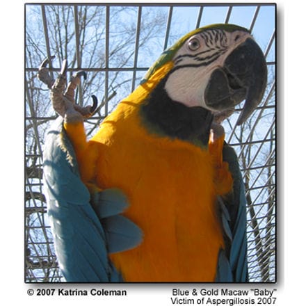
Baby was nine years old and already a master of disguise. He was a blue and gold macaw and loved by everyone. He had a wonderful sense of humor and an insatiable appetite and never even gave a hint of his illness until seven weeks before his death. Louise Bauck, DVM writes, “The first signs are subtle and often missed.” If there were signs, Baby’s were missed. By the time I realized something was wrong it was much too late to save him. Baby was very affectionate and always had a kiss in store for me. He never stopped playing with and tormenting his brother Sammy. He loved hanging around by his toe nails exposing his belly tempting the next passerby to get it.
But his most admirable characteristic was his electric personality and it was present even until the end. I could have never guessed he was so sick. What I wouldn’t give to know then what I know now. I pray that by sharing with you some of what I have learned I might be able to save at least one bird and Baby’s death will not have been in vain.
Most of the following information was learned over the last six weeks of Baby’s life. He was being treated and monitored on a daily basis under the careful and watchful eye of his veterinarian Dr. Sam Vaughn. Dr. Vaughn also made a believer out of me of the importance of an uncontaminated, nutritional diet and clean environment.
The single most memorable piece of advice I was given that I will pass along to you is to never, ever give a parrot a peanut. Every single source of information I have found on this subject is in total agreement. Virginia Caputo, author of Aspergillosis and Jardine’s Parrots says: “Peanuts grow in the ground and are considered to be a common source of aspergillus which can sicken birds.”
“Peanuts are also often contaminated with aflatoxin, a fungal toxin. Aflatoxin is carcinogenic and causes liver damage in birds and other animals. Roasting reduces aflatoxin but does not eliminate it entirely. North American peanut producers are currently working on eliminating contaminated peanuts from their products. Caution is advised when feeding peanuts. Some bird owners, opting to be on the safe side, are eliminating peanuts from their pets’ diet.
Be aware of the possibility of mold growing in all seeds and nuts.”
Aspergillosis is a respiratory fungal infection caused by aspergillus spores that become airborne. Through Dr. Vaughn I also learned spores can be ingested and spread throughout the gastrointestinal tract, and that is how aspergillosis killed Baby. His reluctance to talk was the only sign he presented but I didn’t know it was something I should have been concerned about. Without the silence I may have had a chance to save him. A little known fact to the average bird owner is that acute (sudden and severe) infections often cause a voice to change or disappear completely and aspergillosis is usually not diagnosed until after death. So please, I beg of you, do not wait until you see clinical signs, by then it is usually too late. Take your bird into a qualified avian veterinarian every six (6) months for a check-up. Researchers at The University of Miami have developed a test allowing us to screen parrots for early or mild infections. Subtle changes in bloodwork will tell your vet to investigate further. If I had I wouldn’t be writing this now.
The chronic cases (long term) are somewhat easier to spot. There are symptoms to be seen but they are often associated with other illnesses. A well informed and observant parrot owner has a much better chance of stopping this disease before it consumes your parrot. Some symptoms that can catch your attention are:
- Nasal discharge
- Weight loss (especially if the bird appears to be eating well)
- Diarrhea
- Flakey or delaminating beak
- Unstructured or frayed feathers
- Black edged feathers on the outside of the wings and
- Extreme itchiness and possible mutilation (digging at the skin with beak)
- Loss of or changes in voice
- Neurological problems (including seizures)
Aspergillus is everywhere; it can grow on bread, in rotting vegetation, in materials used to line cages and on living tissue. Every speck of dust and dirt and every batch of corncob bedding have spores of at least one species of Aspergillus. A parrot does not usually succumb to the disease when it has a strong immune system. Over time it has built up antibodies strong enough to wage a proper war against the spores. But, a weakened immune system or ingestion of a huge amount of spores is what causes this disease. Every animal with lungs breathes in thousands of Aspergillus spores every day. The spores cannot grow in the lungs of a healthy body, but a diseased lung can easily become a host. Aspergillosis can consume a parrot with a weakened immune system. Poor nutrition, another illness, anxiety, loneliness, old age, unsanitary conditions or disturbed soil can also bring on this illness. Always keep your parrot’s immune system as healthy as possible. A constant and gradual exposure can create a chronic (long term) infection and the causes are all too commonly found in places you would never expect. Many parrot foods even have the potential to cause a parrot to ingest spores.
Unfortunately, almost all caged birds are eating poor diets, deficient in important vitamins, minerals, amino acids and ample supplies of protein. Stress runs a close race with poor nutrition as being the most common cause of a weakened immune system allowing the Aspergillus spores to grow.;
In every chronic case the slow growing fungus is also very slow to die and recovery can take time. The treatments often last for months or even years. Aspergillosis will never go away on its own.
I’ve listed a few stressors you should be concerned about:
- Spending much of the day in restricted isolation.
- Shipping
- Quarantine
- Overcrowding
- Trauma
- Injury
- Smoke inhalation
- Prolonged antibiotic therapy
- Being a breeder (laying eggs and caring for the young)
A list of possible contaminates that I never would have expected follows. I hope it brings to your attention just how careful you must be.
- PEANUTS, never give a bird a peanut
- Sunflower seeds if their growing season was really wet or the seeds weren’t harvested on time (in essence avoid sunflower seeds since there is no way to detect their history)
- corn cob bedding and other organic matter
- walnut shells
- wood bark (mulch)
- the air during spring planting and fall harvesting on farms
- construction sites where soil is being moved
- damp nesting materials
- potting soil and peat moss, don’t transplant or plant when your birds are around
- wet shavings or other litter
- dried corn
- moldy parrot seed
- fresh fruits and vegetables, if they are not human grade produce and have not been washed with soap and water and a scrub brush
- A warm humid environment, this can speed up the deterioration of nutrients in a parrot’s food and increase the possibility of spores becoming rampant
Remember; “an ounce of prevention is worth a pound of cure.” Once this disease has set in it is very hard to conquer. I have also listed a few simple but critical preventative measures to include in your daily routine.
- Change their water twice a day and any time they have bathed in it or contaminated it.
- Give your parrot fresh food every day.
- Fresh air and exercise are very important, a lack of them can compromise the immune system.
- Remove fresh produce within four hours
- Wash and disinfect cages, toys and perches weekly.
- Boil their water, even bottled or spring water since it is not guaranteed that it doesn’t contain harmful bacteria. A gallon of boiled water kept in the refrigerator will last for days. Just save it for the birds their immune systems are much more delicate than ours.
- Provide good ventilation.
- Provide clean bowls, stainless steel is preferable.
- Provide extra nutrition to your breeders.
- Make sure their nesting materials are clean and dry. Spores can penetrate fresh or incubating eggs and will kill the embryos.
- Eliminate poor ventilation, poor sanitation, dusty conditions and close confinement, these can all increase the chance spores will be inhaled.
My appreciation and thanks are given to Sam Vaughn, BS, DVM, ABVP (AVIAN) of Veterinary Associates Stonefield Louisville KY for taking the time to answer questions about spergillosis, to give advice about preventative measurers and to give me a better chance of giving Sammy a long and healthy life.
Aspergillosis in Birds
Veterinary and Aquatic Services Department, Drs. Foster and Smith, Inc.
What is aspergillosis and what causes it?
Aspergillosis is a respiratory disease of birds caused by the fungus Aspergillus, which is found almost everywhere in the environment.
A. fumigatus is the most common species of the fungus to cause disease, although A. flavus, A. niger, and others can also cause problems. Aspergillus grows readily in warm and moist environments. The microscopic spores of the fungus become airborne, and poor ventilation, poor sanitation, dusty conditions, and close confinement increase the chance the spores will be inhaled.
Usually, the fungus does not cause disease, however, if a bird does not have a healthy immune system, it can cause illness. Predisposing factors include other illnesses, stress, poor nutrition, poor husbandry or unsanitary conditions, another injury to the respiratory system (e.g.; smoke inhalation), and prolonged use of certain medications such as antibiotics or corticosteroids.
The combination of the number of spores in the environment and the presence of predisposing factors determine which birds are most at risk of disease. Aspergillosis appears to be more common in parrots and mynahs than other pet birds.
What are the signs of aspergillosis?
Aspergillosis can follow one of two courses
– acute or chronic. Birds with acute aspergillosis have severe difficulty breathing, decreased or loss of appetite, frequent drinking and urination, cyanosis (a bluish coloration of mucous membranes and/or skin), and even sudden death. The fungus generally affects the trachea, syrinx (= sound-producing vocal organ), and air sacs. The lungs may also be involved. Diagnosis is generally made through a post-mortem examination.
Chronic aspergillosis is much more common, and unfortunately, much more deadly due to its insidious nature. The bird may not become symptomatic until the disease has progressed too far for a cure. The respiratory system is the primary location of infection. White nodules appear and ultimately erode through the tissue, and large numbers of spores enter the bloodstream. The spores then travel throughout the body, infecting multiple organs including kidneys, skin, muscle, gastrointestinal tract, liver, eyes, and brain.
Respiratory symptoms will be the first to occur but will depend on the location of the greatest areas of colonization. Difficulty breathing, rapid breathing and/or exercise intolerance are common. If the syrinx (voice box) is involved, a change in voice, reluctance to talk, or a “click” may occur. Nares may become plugged or you may see a discharge. Eventually, severe respiratory compromise may kill the bird.
Other signs and symptoms will vary, depending on the other organs involved. If any portion of the central nervous system has become involved, the bird may have tremors, an uneven or wobbly gait, seizures, or paralysis. With liver involvement, a green discoloration to the urates may be seen, and the veterinarian may feel an enlarged liver. Generalized, non-specific symptoms can include loss of appetite leading to weight loss, muscle wasting, gout (painful, inflamed joints due to urate deposits), regurgitation, abnormal feces or diarrhea, excessive urination, depression, and lethargy.
Spores can penetrate fresh or incubating eggs and will kill the embryos.
How is aspergillosis diagnosed?
Aspergillosis can be very difficult to diagnose since the signs of disease mimic those of many other illnesses, especially in the chronic form. The veterinarian will need a detailed history of the course of the illness, and an accurate description of the diet and husbandry of the bird. Radiographs, a complete blood count, and a chemistry panel may help support a diagnosis. Endoscopy can be used to view lesions in the syrinx (= sound-producing vocal organ) or trachea, and a sample can be taken for culture and microscopic examination, either of which can confirm a diagnosis. A diagnosis can also be made based on a specific blood test used to detect antibodies to Aspergillus in the blood. Sometimes, however, the test can be falsely negative, especially if the bird’s immune system is suppressed.
How is aspergillosis treated
Surgery may be performed to remove accessible lesions. Antifungal drugs, such as itraconazole, ketoconazole, terbinafine, flucytosine, and amphotericin B, may be administered orally, topically, by injection, or nebulizing, depending upon the drug. Therapy needs to be continued for weeks to months and more than one antifungal drug may be used. Supportive care such as oxygen, supplemental heat, tube feeding, and treatment of underlying conditions are often needed. Unfortunately, the prognosis is always guarded.
How can aspergillosis be prevented?
The importance of good husbandry to prevent outbreaks of aspergillosis cannot be overstated. Keep your bird in a well-ventilated environment. Clean food and water dishes every day. Replace substrate (material lining the cage bottom) regularly. Remove your bird and thoroughly clean cages, toys, perches, etc., at least once a month. Pay attention to good nutrition, offering the right combination of fruits, vegetables, grains and seeds with variety, and only a sprinkling of “treats.” Essentially, you want to do everything you can to alleviate stress in your bird’s life and provide a scrupulously clean environment.
A susceptible bird that has been exposed to Aspergillus may be treated with flucytosine and itraconazole in an attempt to prevent infection.
PetEducation.com – Owned and Operated by Practicing Veterinarians
References and Further Reading
Rupley, AE. Manual of Avian Practice. W.B. Saunders Co. Philadelphia, PA; 1997.
Oglesbee, BL. Mycotic Diseases. Altman, RB; Clubb, SL; Dorrestein, GM; Quesenberry, K. (eds.). Avian Medicine and Surgery. W.B. Saunders Co. Philadelphia, PA; 1997.
Olsen, GH; Orosz, SE. Manual of Avian Medicine. Mosby, Inc. St. Louis, MO; 2000.
Aspergillosis in birds
Aspergillosis is a major cause of morbidity and mortality in birds. It affects birds whether in captive or free-ranging environments, young, mature or geriatric, and whether immunocompetent or immunosuppressed.
Predisposing factors include
- species predilection (turkeys, penguins, raptors, and waterfowl)
- environmental conditions (limited air exchange, exposure to aerosolized toxins resulting in mucosal irritation, and improper temperature and humidity),
- immunosuppression secondary to forced production
- physical exertion (migration), and
- administration of exogenous corticosteroids.
The route of inoculation is inhalation and the lower respiratory tract is the location where Aspergillus spp. tend to initially colonize. A. fumigatus accounts for approximately 95% of the cases and A. flavus is the second most common organism associated with avian infections. – 2005 ISHAM, Medical Mycology, 43, S71/S73 S72 Tell Downloaded At: 18:33 26 August 2007 Clinical signs are usually non-specific (lethargy, inappetance, and anorexia) or can be related to compromise of the respiratory system (rhinitis, change in vocalization, dyspnea).
Diagnostic testing includes:
- Hematologic testing, cytology, fungal culture, histopathology, radiography, and computed tomography.
- Antigen and antibody tests have been reported for use in birds, however their success in properly diagnosing the disease are relatively poor.
The forms of the disease that have been reported in birds include focal aspergilloma, multifocal lesions, and disseminated infections. The target tissues are lungs and air sacs, gross pathologic lesions include caseous nodules or plaques, and massive granulomas with necrotic cores can be appreciated on histopathology. The susceptibility of birds to Aspergillus spp. may be attributed to differences in innate and acquired immunity against this pathogen when compared to mammals.
Anatomic characteristics that might predispose birds to this disease include a lack of an epiglottis for preventing particulate matter from entering the lower respiratory tract, lack of a diaphragm resulting in the inability to produce a strong cough reflex and a limited distribution of pseudostratified ciliated columnar cells through the respiratory tract. Cellular characteristics that might predispose birds to respiratory aspergillosis include a lack of surface macrophages for phagocytizing Aspergillus spp. conidia and dependence on heterophils that use cationic proteins, hydrolases, and lysosymes rather than myeloperoxidase and oxidative mechanisms for killing fungal hyphae [6,7].
The majority of the avian cytokine genes for the Th-1 response have been molecularly characterized; however, cytokines of the Th-2 response still remain elusive despite research efforts. Protective immunity following vaccination for aspergillosis appears promising in turkeys as experimental infections simulating natural disease have been performed and birds appear to confer immunity following vaccination. A unique feature of avian aspergillosis is the presence of reproductive phases of Aspergillus spp. in tissues. This has been reported for the asexual phase of A. fumigatus [8] and both the asexual and sexual phases of A. nidulans [9]. Potential reasons for this finding include the presence of cavernous air sacs, the warm core body temperature of birds (range/38/458C) that might promote conidial germination, and birds’ sensitivity to gliotoxin that might result in tissue necrosis and thus provide a nutrient rich environment.
Given all of these unique characteristics of birds, these animals should no longer be considered as a taxon for which numerous case reports exist, but rather as an interesting and potential experimental animal model for studying this disease. Conclusion Treatment options for resolving Aspergillus infections in animals (i.e. dogs, horses, dolphins, and birds) are still of great need and importance in veterinary medicine. Given the growing population of humans infected with aspergillosis, opportunities to study this disease have significantly increased over the past two decades. These opportunities could benefit both human and veterinary medicine and allow experimental animal models to provide alternative treatment regimens for minimizing morbidity and mortality in the future
On: 26 August 2007
Access Details: Free Access
Publisher: Informa Healthcare
Informa Ltd Registered in England and Wales Registered Number: 1072954
Registered office: Mortimer House, 37-41 Mortimer Street, London W1T 3JH, UK
Medical
Aspergillus and Aspergillosis
by Marilu Anderson, Bird Nutrition and Behavior Consultant
You’ve probably hears of Aspergillus and Aspergillosis, but may not be clear on what these terms mean. First, “Aspergillus” is the name of a common fungus that is naturally present in the environment. For most birds (and people) it causes no problems, but if too many aspergillus organisms are around and your bird has a poor immune system, then the illness “Aspergillosis” sets in. It is often fatal, causing severe respiratory problems. It can be transmitted from birds to humans and vice versa.
Aspergillus spores can be airborne, and are often abundant in corn cob litter. In a damp environment, like we have here in the Northwest, the problem is even worse. I always advise against using this type of litter since it is such a breeding ground for all types of fungus, molds, and bacteria.
Once Aspergillosis has taken hold, the lungs and air sacs fill with large white masses, causing serious breathing problems and further sapping the bird’s energy and immunity. The bird will wheeze, or you’ll hear a clicking sound and often see tail bobbing when the bird’s at rest. Sometimes there is discharge or crustiness around the nostrils. A low grade infection can show as itchiness, frayed feathers, peeling beak or black feather edging onthe wings. There is a blood test for Aspergillosis and it should be part of your bird’s annual checkup.
Treatment is tough – the fungus is hard to kill and because of the weak immune system, there’s often secondary infections as well. Birds on poor diets and living in unsanitary conditions are much more prone to this disease. In dealing with the disease, it’s important to improve the diet, feeding lots of fresh veggies, fruit and using whole food supplements.
Cleanliness also needs to be a top priority, with daily cage cleaning and scrubbing of food and water dishes, as well as perches and toys. Birds on antibiotics for bacterial infections are much more susceptible to Aspergillosis, as well as other fungal and yeast infections. I advise supplying probiotics to birds who are on antibiotics, as well as feeding yogurt and acidophilus. In addition, feed foods rich in Beta Carotene, as Vitamin A is important for good health of the respiratory tract and skin. Yams, carrots, broccoli, red peppers and apricots are all great, as are supplements like wheat grass and spirulina. Many birds really enjoy fresh carrot juice, which is an excellent NATURAL source of Vitamin A. Natural sources are preferable over synthetically produced nutrients, which may not be absorbable and could easily be overdosed).
Boosting your bird’s immune system by supplying a diverse, broad spectrum diet, ensuring adequate rest and daily exercise, and keeping your bird’s cage and supplies scrupulously clean will all help prevent this widespread fungus from grabbing hold in your bird. Don’t forget the importance of regular “well bird” checkups every year, to catch any disease as early as possible, for the best success in treatment. Treating Aspergillosis with antifungal medications needs to be done under direction of an avian vet – it’s not something to try and cure on your own!
Ref.: http://www.yourparrotplace.com
Traditional Treatment:
Aspergillosis is commonly found in decaying vegetable matter. A bird becomes infected by the ingestion or inhalation of mold spores from contaminated foods. The infection causes lesions in the lungs and air sacs and has been reported in many species of birds. Occasionally, outbreaks of the disease cause significant mortality in certain species.
To reduce the risk of this disease:
- Store seed in a dry place to discourage the growth of mold and fungus. Discard any seed that has become wet or moldy.
- Aspergillus fungi are most likely to grow on corn and peanuts. If you live in a warm, humid environment, you may want to avoid feeding these foods.
- It is crucial to not overcrowd birds and to remove fresh foods after a few hours; never leave overnight. No damp, organic matter should be allowed to accumulate in the area the patient(s) is in. Cleanliness is key in both preventing and treating aspergillosis.
Medications: The vet may prescribe Nystatin, Amphotericin B, Ketoconazole or other medications.
Many disease-causing organisms / toxins are transmitted via air and water. If you suspect a disease problem (or if you would like to prevent one), please investigate the possibility of filtering your air and purifying / treating your birds’ drinking water.
Holistic Treatment: Options:
Please note: There is little information available on holistically treating birds and my research took me to resources that pertain to HUMAN patients. The safety for and application to birds has not been ascertained. However, my recommendation is to collect the information and discuss your options with your vet — try to find a vet with additional training in holistic medicine, as regular vets obviously don’t have the knowledge to recommend for or against such treatments.
Remove any possible sources: Moldy food items. Also humidifiers have been mentioned by veterinarians and others as a possible source of mold spores that can cause aspergillosis in birds. Humidifiers need to be properly maintained (as mentioned above). Vets suggest that parrot owners use warm mist humidifiers or vaporizers that boil water prior to releasing it into the air.
Goldenseal Herb: Strong antibiotic properties. It has a long history of use in infections including bacterial, viral, fungal, and parasitic. Staph, strep, E. coli, Vibrio cholera, Giardia lamblia, and even tuberculosis bacterium have proven sensitive to this herb.
Echinacea Herb: Kills a broad range of disease-causing pathogens, including: viruses, bacteria, fungi and protazoa.
Cinnamon Leaf – Essential Oil: Used as an insecticide, emmenagogue, antispasmodic, antibacterial, aphrodisiac and antifungal particularly against Candida and Aspergillis. Indicated for tooth care, blends for vaginitis, respiratory blends for the lungs. Eases colds and breathing difficulties. As an inhalation, it is excellent for exhaustion, feelings of depression and weakness. It is a very effective antiseptic, digestive and anti-rheumatic and is regarded as one of the strongest antiseptic oils. Useful for preventing infectious and contagious diseases. Not recommended for skin care. Traditionally used in clearing warts (I cleared up several warts with this — it worked! Although my skin was sore for a few days. I preferred that to having them cut or frozen off). The leaf oil is relatively non-toxic. Very powerful, should be used with extreme care, could be a skin irritant for certain persons.
References Cited
Abrams, G.A., Paul-Murphy, J., Ramer, J.C. and Murphy, C.J. 2001. Aspergillus blepharitis and dermatitis in a peregrine falcon-gyrfalcon hybrid (Falco peregrinus x Falco rusticolus). Journal of Avian Medicine and Surgery, 15: 114–120.
Arca-Ruibal, B., Wernery, U., Zachariah, R., Bailey, T.A., Di Somma, A., Silvanose, C. and McKinney, P. 2006. Assessment of a commercial sandwich ELISA in the diagnosis of aspergillosis in falcons. The Veterinary Record, 158: 442–444.
Atasever, A. and Gümüssoy, K.S. 2004. Pathological, clinical and mycological findings in experimental aspergillosis infections of starlings. Journal of Veterinary Medicine. A, Physiology, Pathology. Clinical Medicine, 51: 19–22.
Asakura A, Nakagawa S, Masui M, Yasuda J. Immunological studies of aspergillosis in birds. Mycopathol Mycol Appl 19062; 18: 249-56.
Bandera, A., Trabattoni, D., Ferrario, G., Cesari, M., Franzetti, F., Clerici, M. and Gori, A. 2008. Interferon-gamma and granulocyte-macrophage colony stimulating factor therapy in three patients with pulmonary aspergillosis.Infection, 36: 368–373.
Barton, J.T., Daft, B.M., Read, D.H., Kinde, H. and Bickford, A.A. 1992. Tracheal aspergillosis in 6 1/2-week-old chickens caused by Aspergillus flavus. Avian Diseases, 36: 1081–1085.
Bauck , L. , Hillyer , A. & Hoefer , H. 1992 . Rhinitis: case reports . Proceedings of the Annual Conference of the Association of Avian Veterinarians New Orleans LA, USA 134
Beckman, B.J., Howe, C.W., Trampel, D.W., DeBey, M.C., Richard, J.L. and Niyo, Y. 1994. Aspergillus fumigatus keratitis with intraocular invasion in 15-day-old chicks. Avian Diseases, 38: 660–665.
Beernaert, L.A., Baert, K., Marin, P., Chiers, K., De Backer, P., Pasmans, F. and Martel, A. 2009a. Designing voriconazole treatment for racing pigeons: balancing between hepatic enzyme auto induction and toxicity. Medical Mycology, 47: 276–285.
FREE Training to Stop Your Bird's Biting
Get FREE access to Beak School – the only system designed to train your bird. Stop behavioral problems like biting, screaming, feather plucking.
Get the Free TrainingBeernaert, L.A., Pasmans, F., Baert, K., Van Waeyenberghe, L., Chiers, K., Haesebrouck, F. and Martel, A. 2009b. Designing a treatment protocol with voriconazole to eliminate Aspergillus fumigatus from experimentally inoculated pigeons. Veterinary Microbiology, 139: 393–397. doi:doi:10.1016/j.vetmic.2009.06.007
Beernaert, L.A., Pasmans, F., Van Waeyenberghe, L., Dorrestein, G., Verstappen, F.Vercammen, F. 2009c. Avian Aspergillus fumigatus strains resistant to both itraconazole and voriconazole. Antimicrobiol Agents and Chemotherapy, 53: 2199–2201.
Beytut, E. 2007. Immunohistochemical diagnosis of aspergillosis in adult turkeys. Turkish Journal of Veterinary and Animal Sciences, 31: 99–104.
Beytut, E., Özcan, K. and Erginsoy, S. 2004. Immunohistochemical detection of fungal elements in the tissues of goslings with pulmonary and systemic aspergillosis. Acta Veterinaria Hungarica, 52: 71–84.
Bodin E, Gautier L. Note sur une toxine producte par l’Aspergillus fumigatus. Ann Inst Pasteur 1906; 20: 209-24.
Bonar, C.J. and Lewandowski, A.H. 2004. Use of a liposomal formulation of amphotericin B for treating wound aspergillosis in a Goliath heron (Ardea goliath). Journal of Avian Medicine and Surgery, 18: 162–166.
Brown , P.A. & Redig , P.T. 1994 . Aspergillus ELISA: a tool for detection and management. Main conference . In M.J. Kornelsen (Ed.) Proceedings of the Annual Conference of the Association of Avian Veterinarians Reno NV, USA 295
Burhenne, J., Haefeli, W.E., Hess, M. and Scope, A. 2008. Pharmacokinetics, tissue concentrations, and safety of the antifungal agent voriconazole in chickens. Journal of Avian Medicine and Surgery, 22: 199–207.
Cacciuttolo, E., Rossi, G., Nardoni, S., Legrottaglie, R. and Mani, P. 2009. Anatomopathological aspects of avian aspergillosis. Veterinary Research Communications, 33: 521–527.
Carrasco , L. , Lima Jr , J.S. , Halfen , D.C. , Salguero , F.J. , Sanchez-Cordon , P. Becker , G. 2001 . Systemic Aspergillosis in an oiled Magallanic penguin (Spheniscusmagellanicus) . Journal of Veterinary Medicine. B, Infectious Diseases and Veterinary Public Health , 48 , 551 554 .
Carrasco, L., Bautista, M.J., de las Mulas, J.M and Jensen, H.E. 1993. Application of enzyme-immunohistochemistry for the diagnosis of aspergillosis, candidiasis and zygomycosis in three lovebirds. Avian Diseases, 37: 923–927.
Chute HL, O’Meara DC, Barden. A bibliography of avian mycosis (partially annotated). Maine Agr Expt Sta Bull 1962; 655.
Chute HL, O’Meara DC,Tresner HD, Lacombe E. the fungous flora of chickens with infections of p on Avian Health, pp 1-7
Cacciuttolo, E., Rossi, G., Nardoni, S., Legrottaglie, R. and Mani, P. 2009. Anatomopathological aspects of avian aspergillosis. Veterinary Research Communications, 33: 521–527.
Carrasco , L. , Lima Jr , J.S. , Halfen , D.C. , Salguero , F.J. , Sanchez-Cordon , P. & Becker , G. 2001 . Systemic Aspergillosis in an oiled Magallanic penguin (Spheniscusmagellanicus) . Journal of Veterinary Medicine. B, Infectious Diseases and Veterinary Public Health , 48 , 551 554 .
Carrasco, L., Bautista, M.J., de las Mulas, J.M. and Jensen, H.E. 1993. Application of enzyme-immunohistochemistry for the diagnosis of aspergillosis, candidiasis and zygomycosis in three lovebirds. Avian Diseases, 37: 923–927.
Chute HL, O’Meara DC, Barden. A bibliography of avian mycosis (partially annotated). Maine Agr Expt Sta Bull 1962; 655.
Chute HL, O’Meara DC,Tresner HD, Lacombe E. the fungous flora of chickens with infections of the respiratory tract. Am J Vet Res 1956; 17: 763-65.
Chute HL, Witter JF, Rountree JL, O’Meara DCO. The pathology of a fungus infection associated with a capnizing injury. J Am Vet Med Ass 1955; 127: 207-9.
Clark DS, Jones EE, Crowl WB, Ross KF. Aspergillosis in newly hatched chicks. J Am Vet Med Ass 1954; 124: 116-17.
Cray , C. , Watson , T. , Rodriguez , M. & Arheart , K. 2006 . Assessment of aspergillosis diagnostics . In E. Bergman (Ed.) Proceedings of the 27th Annual Conference & Expo of the Association of Avian Veterinarians San Antonio TX, USA 59
Cray, C., Reavill, D., Romagnano, A., Van Sant, F., Champagne, D, .Stevenson, R. 2009a. Galactomannan assay and plasma protein electrophoresis findings in psittacine birds with aspergillosis. Journal of Avian Medicine and Surgery, 23: 125–135.
Cray, C., Watson, T., Rodriguez, M. and Arheart, K.L. 2009b. Application of galactomannan analysis and protein electrophoresis in the diagnosis of aspergillosis in avian species. Journal of Zoo and Wildlife Medicine, 40: 64–70.
Dahlhausen , B. , Abbott , R. & VanOverloop , P. 2004 . Rapid detection of pathogenic Aspergillus species in avian samples by real-time PCR assay: a preliminary report . In E. Bergman (Ed.) . Proceedings of the 25th Annual Conference & Expo of the Association of Avian Veterinarians 37 . New Orleans, LA, USA .
De Herdt, P. 1996. Aspergillose bij papegaaien. Vlaams Diergeneeskundig Tijdschrift, 65: 343–344.
Dhakal,I.P.(2000).Present scenario of poultry farming in Chitawan district of Nepal.Procceding of the Workshop on Avian Health, pp 1-7
Di Somma, A., Bailey, T., Silvanose, C. and Garcia-Martinez, C. 2007. The use of voriconazole for the treatment of aspergillosis in falcons (Falco species). Journal of Avian Medicine and Surgery, 21: 307–316.
Dorrestein, G.M. 1992. De benauwde papegaai. Tijdschrift voor Diergeneeskunde, 117: 55–57.
Eggert MJ, Barnhart JV. A case of egg-borne aspergillosis. J Am Vet Med Ass 1953; 86:781-84.
Fedde, M.R. 1998. Relationship of structure and function of the avian respiratory system to disease susceptibility.Poultry Science, 77: 1130–1138.
Femenia, F., Fontaine, J., Lair-Fulleringer, S., Berkova, N., Huet, D.Towanou, N. 2007. Clinical, mycological and pathological findings in turkeys experimentally infected by Aspergillus fumigatus. Avian Pathology, 36: 213–219.
Flammer , K. & Orosz , S. 2008 . Avian mycoses: managing these difficult diseases . In E. Bergman . Proceedings of the 29th Annual Conference & Expo of the Association of the Avian Veterinarians with the Association of the European College of Avian Medicine and Surgery 153 . Savannah, GA, USA .
Flammer , K. 1993 . An overview of antifungal therapy in birds . In G. Jackson . Proceedings of the Annual Conference of the Association of Avian Veterinarians 1 . Nashville, TN, USA .
Flammer , K. 2006 . Antifungal drug update . In E. Bergman . Proceedings of the 27th Annual Conference & Expo of the Association of Avian Veterinarians 3 . San Antonio, TX, USA .
Forbes, N.A. 1991. Aspergillosis in raptors. The Veterinary Record, 128: 263
Forbes, N.A. 1992. Diagnosis of avian aspergillosis and treatment with itraconazole. The Veterinary Record, 130: 519–520.
Forgacs J, Carll WT. Preliminary mycotoxic studies on hemorrhagic disease in poultry. Vet Med 1955; 50:172.
Heatly , J.J. , Gill , H. , Crandall , L. & Hoerr , F. 2007 . Enilconazole for treatment of raptor aspergillosis . In E. Bergman . Proceedings of the 28th Annual Conference & Expo of the Association of Avian Veterinarians with the Association of the European College of Avian Medicine and Surgery 287 . Providence, RI, USA .
Heatly , J.J. , Gill , H. , Crandall , L. & Hoerr , F. 2007 . Enilconazole for treatment of raptor aspergillosis . In E. Bergman . Proceedings of the 28th Annual Conference & Expo of the Association of Avian Veterinarians with the Association of the European College of Avian Medicine and Surgery 287 . Providence, RI, USA .
Henrici AT. An endotoxin from Aspergillus fumigatus. J Immunol 1939; 36: 319-38.
Hernandez-Divers, S.J. 2002. Endosurgical debridement and diode laser ablation of lung and air sac granulomas in psittacine birds. Journal of Avian Medicine and Surgery, 16: 138–145.
Hofle , U. , Blanco , J.M. , Rodriguez , A. & Vicente , A. 2001 . Atypic aspergillosis—a new threat to the Iberian imperial eagle (Aquila adalberti)? In R. Korbel . Proceedings of the German Veterinary Medical Society, 6th European AAV-DVG Conference of the Association of Avian Veterinarians, 4th Scientific ECAMS Meeting of the European College of Avian Medicine and Surgery 288 . Munich, Germany .
Hoppes, S., Gurfield, N., Flammer, K., Colitz, C. and Fisher, P. 2000. Mycotic keratitis in a blue-fronted Amazon parrot (Amazona aestiva). Journal of Avian Medicine and Surgery, 14: 185–189.
Dhakal, I.P.2000.Present scenario of poultry farming in Chitawan district of Nepal. Procceding of the Workshop on Avian Health, pp 1-7
Ivey, E.S. 2000. Serologic and plasma protein electrophoretic findings in 7 psittacine birds with aspergillosis.Journal of Avian Medicine and Surgery, 14: 103–106.
Jenkins , J. 1991 . Aspergillosis . In Proceedings of the Annual Conference of the Association of Avian Veterinarians 328 . Chicago, IL, USA .
Jensen, H.E., Christensen, J.P., Bisgaard, M. and Nielsen, O.L. 1997. Immunohistochemistry for the diagnosis of aspergillosis in turkey poults. Avian Pathology, 26: 5–18.
Jones, M.P. and Orosz, S.E. 2000. The diagnosis of aspergillosis in birds. Seminars in Avian and Exotic Pet Medicine, 9: 52–58.
Jones, M.P., Orosz, S.E., Cox, S.K. and Frazier, D.L. 2000. Pharmacokinetic disposition of itraconazole in red-tailed hawks (Buteo jamaicensis). Journal of Avian Medicine and Surgery, 14: 15–22.
Joseph , V. , Pappagianis , D. & Reavill , D.R. 1994 . Clotrimazole nebulization for the treatment of respiratory aspergillosis . In M.J. Kornelsen . Proceedings of the Annual Conference of the Association of Avian Veterinarians 301 . Reno, NV, USA .
Joseph, V. 2000. Aspergillosis in raptors. Seminars in Avian and Exotic Pet Medicine, 9: 66–74.
Julian, R.J. and Goryo, M. 1990. Pulmonary aspergillosis causing right ventricular failure and ascites in meat-type chickens. Avian Pathology, 19: 643–654.
Jung, K., Kim, Y., Lee, H. and Kim, J.T. 2009. Aspergillus fumigatus infection in two wild Eurasian black vultures (Aegypius monachus Linnaeus) with carbofuran insecticide poisoning: a case report. The Veterinary Journal, 179: 307–312.
Kaufman, L., Standard, P.G., Jalbert, M. and Kraft, D.E. 1997. Immunohistologic identification of Aspergillus spp. and other hyaline fungi by using polyclonal fluorescent antibodies. Journal of Clinical Microbiology, 35: 2206–2209.
Khosravi, A.R., Shokri, H., Ziglari, T., Naeini, A.R., Mousavi, Z. and Hashemi, H. 2008. Outbreak of severe disseminated aspergillosis in a flock of ostrich (Struthio camelus). Mycoses, 51: 557–559.
Le Loch , G. , Deville , M. , Risi , E. , Bretagne , S. & Guillot , J. 2005 Evaluation of the serological test platelia spergillus for the diagnosis of aspergillosis . In T. Bailey , J. Chitty , N. Harcourt-Brown & J. Samour .Proceedings of the 8th European Conference of the Association of Avian Veterinarians, 6th Scientific ECAMS Meeting of the European College of Avian Medicine and Surgery 260 . Arles, France .
Low, M., Berggen, A., Morgan, K.J. and Alley, M.R. 2005. Aspergillosis in a North Island robin (Petroica longipes).New Zealand Veterinary Journal, 53: 462–464.
Lucet A. Experimental and clinical study of Aspergillus fumigatus. Vet J 1897; 44: 215-17, 285-88, 292-94; 45: 226-31, 301-4.
Lumeij, J.T., Gorgevska, D. and Woestenborghs, R. 1995. Plasma and tissue concentrations of itraconazole in racing pigeons (Columba livia domestica). Journal of Avian Medicine and Surgery, 9: 32–35.
Maina, J.N. 2002. Some recent advances on the study and understanding of the functional design of the avian lung: morphological and morphometric perspectives. Biological Reviews of the Cambridge Philosophical Society, 77: 97–152.
Marks, S.L., Stauber, E.H. and Ernstrom, S.B. 1994. Aspergillosis in an ostrich. Journal of the American Veterinary Medical Association, 204: 784–785.
McMillan, M.C. and Petrak, M.L. 1989. Retrospective study of aspergillosis in pet birds. Journal of the Association of Avian Veterinarians, 3: 211–215.
Meredith , A. 1997 . Prophylactic administration of itraconazole for the control of aspergillosis in gentoo penguins (Pygoscelis papua) . In The 4th Conference of the European Committee of the Association of Avian Veterinarians 227 . London, UK .
Nardoni, S., Ceccherelli, R., Rossi, G. and Mancianti, F. 2006. Aspergillosis in Larus cachinnans micaellis: survey of eight cases. Mycopathologia, 161: 317–321.
Oglesbee, B.L. 1997. “Mycotic diseases”. In Avian Medicine and Surgery, 1st edn, Edited by: Altman, R.B. 323–361. Philadelphia, PA: W.B. Saunders Company.
O’Meara DC, Chute HL. Aspergillosis experimentally bproduced in hatching chicks. Avian Diseases 1959; 3: 404-6.
Orosz, S.E. and Frazier, D.L. 1995. Antifungal agents: a review of their pharmacology and therapeutic indications.Journal of Avian Medicine and Surgery, 9: 8–18.
Orosz, S.E., Frazier, D.L., Schroeder, E.C., Cox, S.K., Schaeffer, D.O., Doss, S. and Morris, P.J. 1996. Pharmacokinetic properties of itraconazole in blue-fronted Amazon parrots (Amazona aestiva aestiva). Journal of Avian Medicine and Surgery, 10: 168–173.
Peden, W.M. and Rhoades, K.R. 1992. Pathogenicity differences of multiple isolates of Aspergillus fumigatus in turkeys. Avian Diseases, 36: 537–542.
Perelman , B. 1993 . Evaluation of azole anti-mycotic agents using an experimental model of aspergillosis in turkey poults . In Proceedings of the European Conference on Avian Medicine and Surgery 120 . Utrecht, The Netherlands .
FREE Training to Stop Your Bird's Biting
Get FREE access to Beak School – the only system designed to train your bird. Stop behavioral problems like biting, screaming, feather plucking.
Get the Free TrainingPerelman, B. and Kuttin, E.S. 1992. Aspergillosis in ostriches. Avian Pathology, 21: 159–163.
Pericard , J.M. 2005 . Clinical assessment on the use of fluconazole per os in 24 African grey parrots (Psittacus erithacus): acceptance, side effects and efficiency . In T. Bailey , J. Chitty , N. Harcourt-Brown & J. Samour . Proceedings of the 8th European Conference of the Association of Avian Veterinarians, 6th Scientific ECAMS Meeting of the European College of Avian Medicine and Surgery 222 . Arles, France .
Phalen, D.N. 2000. Respiratory medicine of cage and aviary birds. Veterinary Clinics of North America: Exotic Animal Practice, 3: 423–452.
Redig , P.T. , Orosz , S. & Cray , C. 1997 . The ELISA as a management guide for aspergillosis in raptors . In M. Doolen (Ed.). In Proceedings of the Annual Conference & Expo of the Association of Avian Veterinarians 99 . Reno, NV, USA .
Redig , P.T. 1994 . Diagnosis of avian aspergillosis Kornelsen M.J. Main Conference Proceedings of the Annual Conference of the Association of Avian Veterinarians 355 . Reno, NV, USA .
Redig, P.T. and Duke, G.E. 1985. Comparative pharmacokinetics of antifungal drugs in domestic turkeys, red-tailed hawks, broad-winged hawks, and great-horned owls. Avian Diseases, 29: 649–661.
Reidarson , T.H. & McBain , J. 1995 . Serum protein electrophoresis and Aspergillus antibody titers as an aid to diagnosis of aspergillosis in penguins Kornelsen M.J. Proceedings of the Annual Conference & Expo of the Association of Avian Veterinarians 61 . Philadelphia, PA, USA .
Richard, J.L. and Thurston, J.R. 1983. Rapid hematogenous dissemination of Aspergillus fumigatus and A. flavusspores in turkey poults following aerosol exposure. Avian Diseases, 27: 1025–1033.
Richard, J.L., Dvorak, T.J. and Ross, P.F. 1996. Natural occurrence of gliotoxin in turkeys infected with Aspergillus fumigatus, Fresenius. Mycopathologia, 134: 167–170.
Richard, J.L., Peden, W.M. and Sacks, J.M. 1991. Effects of adjuvant-augmented germling vaccines in turkey poults challenged with Aspergillus fumigatus. Avian Diseases, 35: 93–99.
FREE Training to Stop Your Bird's Biting
Get FREE access to Beak School – the only system designed to train your bird. Stop behavioral problems like biting, screaming, feather plucking.
Get the Free TrainingRichard, J.L., Peden, W.M. and Williams, P.P. 1994. Gliotoxin inhibits transformation and is cytotoxic to turkey peripheral blood lymphocytes. Mycopathologia, 126: 109–114.
Schmidt, A. 2002. Animal models of aspergillosis—also useful for vaccination strategies. Mycoses, 45: 38–40.
Schmidt, V., Demiraj, F., Di Somma, A., Bailey, T., Ungemach, F.R. and Krautwald-Junghanns, M.E. 2007. Plasma concentrations of voriconazole in falcons. The Veterinary Record, 161: 265–268.
Scope , A. , Burhenne , J. , Haefel , W.E. & Hess , M. 2007 . Species dependent differences and evaluation of possible influences on the enteral absorption of voriconazole in birds . In Proceedings of the 9th European Conference of the Association of Avian Veterinarians, 7th Scientific ECAMS Meeting of the European College of Avian Medicine and Surgery 236 . Zürich, Switzerland .
Silvanose , C.D. , Bailey , T.A. & Di Somma , A. 2009 . Invitro susceptibility of Aspergillus sp. isolated from the respiratory tract of falcons Martel A. Proceedings of the 10th European Conference of the Association of Avian Veterinarians, 8th ECAMS Scientific Meeting of the Association of the European College of Avian Medicine and Surgery 355 . Antwerp, Belgium .
Silvanose, C.D., Bailey, T.A. and Di Somma, A. 2006. Susceptibility of fungi isolated from the respiratory tract of falcons to amphotericin B, itraconazole and voriconazole. The Veterinary Record, 159: 282–284.
Snelders, E., van der Lee, H.A.L., Kuijpers, J., Rijs, A.J.M.M., Varga, J.Samson, R.A. 2008. Emergence of azole resistance in Aspergillus fumigatus and spread of a single resistance mechanism. PLoS Medicine, 5: 1629–1637.
Soike, D., Köhler, B. and Albrecht, K. 1999. A circovirus-like infection in geese related to a runting syndrome. Avian Pathology, 28: 199–202.
Suedmeyer, W.K., Bermudez, A.J. and Fales, W.H. 2002. Treatment of epidermal cysts associated with Aspergillus fumigatus and Alternaria species in a silky bantam chicken. Journal of Avian Medicine and Surgery, 16: 133–137.
Taylor , M. 1993 . A new endoscopic system for the collection of diagnostic specimens in the bird Jackson G.Proceedings of the Annual Conference of the Association of Avian Veterinarians 83 . Nashville, TN, USA .
Tell, L.A. 2005. Aspergillosis in mammals and birds: impact on veterinary medicine. Medical Mycology Supplement, 1: S7–S73.
Tell, L.A., Clemons, K.V., Kline, Y., Woods, L., Kass, P.H., Martinez, M. and Stevens, D.A. 2009. Efficacy of voriconazole in Japanese quail (Coturnix japonica) experimentally infected with Aspergillus fumigatus. Medical Mycology, 48: 234–244.
Thom C, Church MB. The Aspergilli . Williams & Wilkins Co, Baltimore. 1926.
Toth, T.E. 2000. Nonspecific cellular defense of the avian respiratory system: a review. Developmental and Comparative Immunology, 24: 121–139.
Toth, T.E. and Siegel, P.B. 1986. Cellular defense of the avian respiratory tract: paucity of free-residing macrophages in the normal chicken. Avian Diseases, 30: 67–75.
Tsai, S.S., Park, J.H., Hirai, K. and Itakura, C. 1992. Aspergillosis and candidiasis in psittacine and passeriforme birds with particular reference to nasal lesions. Avian Pathology, 21: 699–709.
Van Heelsbergen T. Handbuch der Gefluegelkrankheiten und der Gefluegelzucht. Ferdinand Enke, Stuttgart 1929; Pp. 312-22.
Van Waeyenberghe , L. , Pasmans , F. , Beernaert , L.A. , Haesebrouck , F. & Martel , A. 2009 . Interaction of the avian macrophage with Aspergillus fumigatus: to let live or let die . In A. Martel (Ed.) . Proceedings of the 10th European Conference of the Association of Avian Veterinarians, 8th ECAMS Scientific Meeting of the Association of the European College of Avian Medicine and Surgery 375 . Antwerp, Belgium .
Vanderheyden , N. 1993 . Aspergillosis in psittacine chicks Jackson G. Proceedings of the Annual Conference of the Association of Avian Veterinarians 207 . Nashville, TN, USA .
Verstappen, F.A.L.M. and Dorrestein, G.M. 2005. Aspergillosis in Amazon parrots after corticosteroid therapy for smoke-inhalation injury. Journal of Avian Medicine and Surgery, 19: 138–141.
Westerhof, I. 1995. Treatment of tracheal obstruction in psittacine birds using a suction technique: a retrospective study of 19 birds. Journal of Avian Medicine and Surgery, 9: 45–49.
Xavier , M.O. 2008 . A review of aspergillosis in penguins . Available online at http://www.aspergillus.org.uk/secure/articles/aspergillo.pdf accessed Jan. 2012 .
Young, E.A., Cornish, T.E. and Little, S.E. 1998. Concomitant mycotic and verminous pneumonia in a Blue Jay from Georgia. Journal of Wildlife Diseases, 34: 625–628.
Information contained on this website is provided as general reference only. For application to specific circumstances, professional advice should be sought.
Please Note: The articles or images on this page are the sole property of the authors or photographers. Please contact them directly with respect to any copyright or licensing questions. Thank you.