Introduction
Avian Bornaviral Ganglioneuritis (ABG) is an immune-mediated disease and is
responsible for what has been previously known as Proventricular Dilatation Disease
(PDD). As many as one-third of the avian population in captivity and in the wild test
positive for the Avian Bornavirus (ABV). Although captive psittacines are the most
impacted, other species also test positive for ABV. ABV does not appear to be zoonotic; humans and animals do not contract this infection from pet birds. It causes disease in the class Aves which includes many species of birds. The Avian Bornavirus may be transmitted both horizontally and vertically. Treatment protocols and proper care are extending the lives of many birds affected with Avian Bornaviral disease. Without treatment, it is usually fatal.
Much of the information available about Avian Bornaviral Disease (ABVD) is now out-of-date. In an attempt to clarify the nature of this disease, researchers now refer to PDD as Avian Ganglioneuritis (AG). The ganglioneuritis may be caused by the Avian
Bornavirus, or it may be caused by another illness. If the bird has not tested positive by molecular diagnostic testing, then the term Avian Ganglioneuritis (AG) should be
applied to the illness, and it only affects the Gastrointestinal System. If it has tested
positive, the term Avian Bornaviral Ganglioneuritis (ABG) is used and it affects both the GI track and the Central Nervous System (CNS). The treatment is the same for both. It is important to have the bird tested for the Avian Bornavirus so the owner can know if the illness is caused by the Avian Bornavirus or another disease state. If the bird does not test positive for the virus, other tests are in order to find the cause.
The term, “Avian Ganglioneuritis” better describes the ongoing disease process and
shifts the emphasis from the proventriculus (gastric stomach) to all the affected organs and systems [4]. This paper’s focus is on the ganglioneuritis and CNS symptoms caused by the Avian Bornavirus, but the reader must keep in mind that Avian Ganglioneuritis can be caused by any one of a number of other illnesses.
FREE Training to Stop Your Bird's Biting
Get FREE access to Beak School – the only system designed to train your bird. Stop behavioral problems like biting, screaming, feather plucking.
Get the Free TrainingThe following is a statement by Dr. Bob Dahlhausen with the most current information existing to date. Dr. Dahlhausen is a foremost researcher into this disease and is the veterinarian who first used Celebrex (celecoxib) in his research and with patients.
Avian Ganglioneuritis (Avian Borna Virus [ABV] and Proventricular Dilatation
Disease [PDD])
“Some people think the ABV is just like any other virus: you get it, get sick, and get over it. But ABV is not like that. ABV infection rates are wide-scale. A survey of aviaries in Europe of 1442 birds showed a 23% infection rate. This is similar to the infection rate of wild birds. The average is 30% in almost all aviaries. It is everywhere. One third of birds will contract it.
The dilemma is that a lot of birds have classic PDD signs yet when tested do not have
the Borna virus. What is going on with the pathogenesis? We now know that PDD is an autoimmune disease similar to Guillain-Barre Syndrome.
Throughout the whole nervous system of the bird there are these little proteins called
‘gangliosides’. There are about 50 different ones, and they vary in location throughout
the nervous system of the bird. The virus can expose these proteins, and once they are
exposed, the host’s immune system will mount a cross-reaction to the nerve ganglia
proteins. This causes an autoimmune reaction and disease.
The true role of the Bornavirus is that it can directly or indirectly damage the nerves that cause the gangliosides to be exposed. Then there is another mechanism. It is called ‘antigen mimicry’. That means that infections with agents such as herpesvirus,
Campylobacter and Chlamydophila are so similar to the ganglioside proteins that if the bird’s body mounts an immune response to any of these agents, the immune response will cross-reference to the bird’s nervous system. So we can have infections with these agents, and the bird can show symptoms of PDD when there’s no Bornavirus present.
So many vets and owners think that the Bornavirus infects the bird, the bird gets sick
and gets the disease. The Bornavirus is a cause of PDD signs, but it’s not the only
cause. The dilemma is that birds can have classic PDD signs yet be ABV negative. In
Italy, the researchers have a theory: Can we sensitize the bird against the ganglioside
proteins and produce PDD? Can ganglioside sensitization cause the PDD signs? They
took 8 cockatiels. Three were given ganglioside proteins orally, and 3 were given the
gangliosides by injection. One month later 100% of injected birds and 33% of the birds that received the oral gangliosides developed Central Nervous system (CNS) and gastrointestinal (GI) signs compatible with PDD. The histopathology was identical to PDD. They caused PDD without the presence of ABV. This leads us to conclude that any infection that exposes nerve gangliosides to the host’s immune system and elicits a suitable immune response can produce disease that is compatible with PDD. You can’t tell the difference; many things can cause it. It is true that ABV infection is a cause of PDD, but it’s not the only cause.
When we talk about testing, 1/3 of birds will test positive. A lot of people think that if a bird tests positive, it is a fatal disease. It is going to waste away and die, and it’s very contagious. This is not the case. The majority of birds that are positive are disease-free. The virus is very unstable in the environment. It loses infectivity within 8 hours. It can’t live in the environment, so it does not easily go from bird to bird. In fact, we believe the major transmission is through the egg; the birds are born with this infection. It is not highly contagious via normal routes of exposure. The Italian researchers have taken live birds and put the virus in the nares (nostrils) to simulate inhalation of it and not a single one of those birds became infected.”
Anti-ganglioside Antibody Assay
“This is a new test we are using for ABV/PDD. Testing for the virus is pretty
meaningless with traditional testing methods. There’s a 30% chance your bird will get
sick. What we have now is the Anti-ganglioside Antibody Assay. It’s an ELIZA
(molecular diagnostic blood) test. It measures the antibodies—the immune response of the bird against the ganglioside proteins. This is a much more accurate test. If you have a bird with seizures or is wasting and not digesting well, or wobbly, having ataxia (difficulty balancing and controlling its muscles), this test will show if it has
ganglioneuritis, and we can treat it. Over 98% of birds that were showing clinical
disease symptoms like PDD tested positive on that assay. We’re not testing for Avian
Bornavirus anymore. We are testing for the ganglioside antibodies. We now have really good ways of treating birds. There is a lot we can do for them. PDD, or the wasted bird, is the worst-case scenario. We see a lot of subtle disease before that and varying degrees of disease. We can treat them so they can live a nice, long, healthy life.”
1. Worldwide Distribution and Impact of Avian Bornaviral Ganglioneuritis
The disease, originally known as “Macaw Wasting Disease”, was initially reported in the late 1970’s in the U.S. and Europe. The term, “Proventricular Dilatation Disease”, was given to the disease in a 1983 report describing “impaction, dilation, and degeneration of the proventriculus” [4]. As of 2009, verified cases had been reported in as many as 80 psittacine and non-psittacine species, both captive and wild. Since then, the Avian Bornavirus has been proven to be a cause of Avian Bornaviral Ganglioneuritis and has been detected in birds around the world. This disease “now presents a serious threat to both captive propagation and conservation efforts for endangered psittacines such as the Spix macaw” [4].
2. Defining Avian Bornaviral Ganglioneuritis
The Avian Bornavirus is an enveloped, negative-stranded, ribonucleic acid (RNA) virus. The virus causes Avian Bornaviral Ganglioneuritis which is a fatal, inflammatory wasting disease affecting mostly birds in the psittacine family (Order Psittaciformes) [1]. It is a disorder which impacts the nervous system and causes gastrointestinal and
neurological dysfunction [2]. In pathology terms, it is described as a “non-suppurative,
lymphocytic-plasmacytic ganglioneuritis of the nerve plexi of the crop, proventriculus,
ventriculus, and duodenum and peripheral and central nervous systems” [2,18].
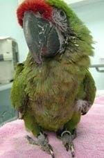
Figure 1. A macaw suffering from Avian Bornaviral Ganglioneuritis. Note the poor
condition of the beak and feathers and depressed stance (image courtesy of R.
Dahlhausen, used with permission).
3. Etiology and Pathogenesis of Avian Bornaviral Disease (ABD)
In the 1970’s, when the great influx of birds from other countries began, captive birds
showed clinical signs of ganglioneuritis, and researchers suspected a viral etiology [2]. In 2010, viral inclusion bodies and enveloped particles were discovered in the myenteric plexus (an arrangement of nerve fibers and neuron cells that are situated within the muscular tissue layer. This layer encloses the esophagus, stomach, and intestines and celiac ganglion of infected birds) [15]. Independent researchers provided confirmation that the etiological agent and pathogenesis of ABG were related to Bornavirus. This novel genome was named “Avian Bornavirus” [1,15]. The connection between Borna Disease Virus (BDV) and Avian Bornaviral Disease was established using qPCR (Realtime Polymerase Chain Reaction) [2].
Avian Bornavirus has been found in tissues of the brain, proventriculus, plasma, crop,
ventriculus, duodenum, liver, lung, kidney, spleen, retina, cerebrum, cerebellum and
adrenal glands of infected birds [2]. Gancz was able to transmit the infection by
inoculating birds with tissue from ABV-positive birds. Not all developed signs, but all
were found to have the characteristic lymphoplasmacytic infiltrates in the myenteric
ganglia (collection of nerve groups within the intestinal tract) and variable degrees of
lesions in the brain and spinal cord [15].
ABG had previously been regarded as carrying a high-mortality but low-infection risk. As of a few years ago, it has been shown to occur much more frequently but involve a “low incidence of clinical disease and a much lower incidence of severe disease” [12]. New treatment protocols have moved this disease to a more chronic state and one which often responds to treatment. Nevertheless, an infected bird will never eliminate the viral infection [2].
3.1 The Parent Disease: Borna Disease Virus
Borna Disease Virus was originally discovered in cavalry horses in Borna, Germany in 1885. Since that time, it has become known as a “neurological disease of a wide range of animal species and possibly humans” [2,4].
Most viruses spread by cell-to-cell contact, first invading and destroying the host cell,
then moving on to infect more cells. Borna Disease Virus, however, uses the “nuclear
compartment of the host cells in which infectious ribonucleoproteins are present for
transcription and replication” [15]. The virus does not destroy the cell, so infected cells suffer very little damage. Additionally, the virus has developed several methods to avoid being recognized by the host immune system [2,3]. “This is a detection-evasion strategy which allows the molecules to go unrecognized by the cytosolic RNA sensor that triggers the host’s innate immune response” [15]. To accomplish this, the virus must suppress apoptosis (natural cell death.) The result is continual and lifelong infection for both Bornavirus and Avian Bornavirus. ABG is now known to be an immune-mediated disease [15].
Avian Bornavirus is different from Bornavirus —it does not grow in mammalian cell
lines; therefore, it is not thought to infect humans or animals [2]. BDV attacks are limited to the central nervous system; however, ABV affects the innervation of multiple organs: the brain, GI tract, liver, kidneys, heart, and lungs, as well as in the peripheral blood vessels [3]. ABV is a neurotropic virus: It attacks the central, peripheral and autonomic nervous systems. There is a wide range of signs from the infection. In some birds, only slight changes in behavior are noted, but in others, the birds suffer severe neurological disease which results in fatal infections [2,4].
4. Infection Rates
Practitioners have been reporting high numbers of birds testing positive for ABV. Lierz
detected ABV RNA in 45.8% of normal birds [10]. In a 2010 study of molecular samples from 791 psittacines, ABV RNA was discovered in 34.3% of the samples. In a later study, 100+ psittacines living in a stressful shelter situation were tested, and 52% of this population tested positive for ABV-specific RNA. In some densely populated shelters, even higher numbers of birds have tested positive [3].
5. The Avian Bornavirus and its Genotypes
The term “genotype” refers to the genetic makeup of an individual virus. As of 2017, the family Bornaviridae contained a diverse viral group of 15 different genotypes. Of those, genotypes 2 and 4 are frequently found in psittacine species [18]. Viral shedding is intermittent; therefore, a sample taken when the virus is not being shed will test negative for the virus. Additionally, the virus is very unstable and will degrade quickly, producing a negative test result. Finally, not all birds with Avian Ganglioneuritis test positive for Avian Bornavirus [2].
6. Detection of Avian Bornaviral Disease
Clinical signs vary from infrequent mild episodes to sudden and acute illness [10]. A bird may experience just some of the signs, but not necessarily all. Some birds becomesymptomatic years or decades after becoming ABV-infected, and some never show signs at all but may continue to shed the virus and thus infect other birds [4]. Healthy birds can experience viremia (the presence of the virus in the blood) due to ABV without becoming clinically ill [2]. In fact, most of the ABV-positive birds do not display clinical disease [12].
6.1 Challenges Encountered When Testing for Avian Bornaviral Infections
- Lymphoplasmacytic ganglioneuritis (infected nerve groups), pathological lesions, or a similar disease state may be caused by other infectious agents [1,2]. One other virus, paramyxovirus-1, may cause the same ganglioneuritis signs as ABV disease [2].
- Some psittacine species are more vulnerable to specific genotypes than others.
- The various ABV genotypes may each produce distinctive and specific clinical signs. This may explain why some birds develop certain signs and others experience different signs, and why some birds, when exposed, do not contract the virus at all.
- There are minor variations, but they all produce the same general pathology [2].
- Some reported primer sequences used in ABV Reverse-Transcription Polymerase Chain Reaction (RT-PCR) testing do not detect all identified ABV genotypes [2].
- “While the virus can be detected in cloacal swabs from both clinically and nonclinically infected birds, cloacal swab samples are not consistently positive in affected birds, possibly due to the high level of RNA enzymes in fecal material that can destroy viral RNA and the inconsistent shedding of the virus” [2].
- Other destructive factors may be present, like bacteria, enzymes, or other contaminants found in the feces [4]. These artifacts can result in a false negative test result.
7. Species Affected by the Avian Bornavirus
Psittacines appear to be the most affected by the Avian Bornavirus; as a result, almost
all of the studies have concentrated on the captive psittacine population. The species
with the highest number of positive results were cockatoos, Amazons, Eclectus, African greys, and macaws. Quakers and lovebirds have been minimally represented. Those with the lowest number were cockatiels, budgerigars, pionus and conures [4].
7.1 ABV in Wild Psittacines and Non-psittacine Species
Investigations of ABV infection in wild psittacines had not been conducted until recently. In 2011, more than 80 free-ranging, clinically healthy psittacines were tested for ABV. The birds were located in Brazil and belonged to seven different species. The results revealed that 33% tested positive for genotype 4, and 50% of the birds possessed antibodies to the virus [7]. Avian species other than psittacines are vulnerable to ABV, specifically waterfowl. In 2011, researchers tested Mute Swans, Snow Geese, Ross’ Geese, and Greater White-fronted Geese. ABV was detected in all but the Whitefronted Geese. Of the ducks tested, 11% of ducks and 13% of gulls tested positive. Acute encephalitis (inflammation of the brain) was found in geese and swans. Bald Eagles have been found with the virus; it is assumed that raptors could be vulnerable through predation [3,13]. Canaries (order Passeriformes) have been found to be infected with ABV. One canary showed signs of apathy just three days before it succumbed to the virus [11]. The lesions found in it were similar to the lesions found in the psittacine birds with ABV disease. Both neural and extraneural tissues contained Bornaviral antigens (a substance that evokes an immune response from the body) [6]. A second canary’s illness was chronic. The symptoms included prolonged depression, CNS (Central Nervous System) signs, and visual impairment with chorioretinitis (ocular inflammation)” [12]. Cortical blindness may occur in canaries. It often responds to treatment (R. Dahlhausen, personal communication).
8. Periods of Vulnerability
8.1 Young Birds
Unweaned birds are more vulnerable to ABV infection than adults. The estimated
incubation period is between two and four weeks or longer [6]. Avian Bornavirus’
ordinarily long incubation period is shorter for the immunocompromised or very young
with incomplete immune system development [13].
8.2 The Breeding Season
Hormonal and reproductive activity often give rise to new cases and relapses of
previous cases. The disease cycles from active to dormant states, and the stresses of
this increased hormonal state depress the immune system, allowing clinical disease to
flare up [2]. One clinician has treated a patient who had experienced severe relapses of gastrointestinal symptoms during four consecutive breeding seasons [2]. Each
consecutive relapse diminishes the bird’s ability to fight the disease and further
damages the organs.
9. Development of Signs
In some naturally occurring flocks, ABG is described as a “sporadic disease” [2]. Even
single pet birds, housed in the same environment for many years, have been diagnosed with the disease. The clinician should not automatically assume a bird is afflicted with ABG simply because it tests positive for ABV. It might take years before an ABV positive bird develops signs of ABG, and some never become symptomatic. It is not known if or when the bird will become symptomatic or what will trigger the onset of disease [6].
9.1 Factors in the Bird’s Reaction to the Presence of the Virus
ABV disease is “defined by the absence or presence of antigenemia” (having an antigen in the blood) [2]. Each of the following factors will influence or change the way the bird reacts to the virus, and these signs differ from case to case. R. Dahlhausen attributes the development of clinical disease to the following predisposing factors:
- Genetics, age, host species, and ABV genotype involved.
- Severity of the disease, distribution of lesions, and involvement of affected organ systems [4].
- The developmental competency or compromised condition of the host’s immune system [2].
- Stress due to malnutrition, concurrent disease, reproductive activity, and improper husbandry. Stress is generally accepted as the primary predisposing factor in the activation or recurrence of ABG [2].
10. The Avian Bornavirus’ Effect on the Systems of the Body
Avian Bornavirus primarily affects the CNS and nerves of the gastrointestinal tract [4].
Birds may exhibit only GI or CNS signs, but some experience both, and the virus usually affects both, even if they are not initially obvious.
In a 2015 survey conducted by Dahlhausen and Orosz, 66% of birds exhibited CNS
signs, 22% experienced GI tract signs, 9% displayed feather picking and mutilation, and 9% suffered acute death [4].
10.1 Gastrointestinal Signs of Avian Bornaviral Disease
“Avian Bornaviral Ganglioneuritis is a persistent inflammation of the networks of nervous tissues due to the invasion of lymphocytes and plasmacytes into the nerve ganglia of organs of the upper digestive tract” [2,14,18]. Affected birds are unable to digest or absorb their dietary nutrients properly. Sometimes, secondary bacterial or fungal infections of the GI tract result from poor motility.
The GI signs indicate pathology of the vagus nerve (Cranial Nerve X) [4]. The GI
symptoms develop from the disruption of innervation to the vagus nerve [12]. This nerve “supplies the proximal (upper) portion of the intestinal tract—from the crop,
proventriculus and ventriculus to the duodenum and the heart” [12]. When the vagus
nerve malfunctions, the normal GI motility is altered, and the wall of the GI tract
becomes thin [12]. This can result in the rupture of the proventriculus and immediate,
very painful, death.
“Finding the infected nerve ganglia confirms that the individual is suffering from Avian
Ganglioneuritis” [12]. Clinical signs include:
FREE Training to Stop Your Bird's Biting
Get FREE access to Beak School – the only system designed to train your bird. Stop behavioral problems like biting, screaming, feather plucking.
Get the Free Training- Depression
- Weight loss and abdominal enlargement
- Muscular and neurogenic atrophy
- Crop stasis and regurgitation- Polyuria (excessive urination)
- Weakness and loss of body condition
- Passage of undigested food in the feces
- Poor absorption and impaired GI tract transit
- Polyphagia (excessive eating) [12].
As long as the intestinal tract is distended, the proventriculus and ventriculus will not become fully emptied; motility will be inhibited, and gastrointestinal stasis may result [4].

Figure 2. Droppings containing undigested food (image courtesy of
www.vetexotic.theclinics.com, used with permission).

Figure 3. Displacement of the organs due to dilated proventriculus. “This bird was very thin with a very prominent keel bone and pectoral muscle atrophy. The crop and
proventriculus were markedly dilated and filled with soft oats. The proventriculus filled 80% of the cranial coelom and displaced the liver dorsally and to the right. The
ventriculus was displaced caudally and displaced the intestines dorsally. The intestines contained a small amount of brown liquid.” (vetpath.wordpress.com, used with permission).

Figure 4. Necropsy photo of a crop containing undigested foods. Whole seeds can be
seen in this picture (image courtesy of Avian Genetics, used with permission).
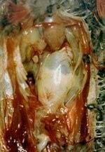
Figure 5. Necropsy photos showing the displacement of organs caused by dilated
proventriculus and ventriculus. The large, pale, center organ (the proventriculus) and
the smaller, yellowish extension of it (the ventriculus) are displacing the lungs, kidneys, heart, and liver (image courtesy of R. Dahlhausen, used with permission).

Figure 6. A necropsy photo displaying the wasting effect of the disease in a cockatoo
(image courtesy of R. Dahlhausen, used with permission).
10.2 Neurological Signs of Avian Bornaviral Disease
CNS lesions are usually found in the cerebrum or cerebellum. Disruptions of the cell layers within the cerebellum produce disorders in fine-muscle movement and equilibrium [4]. The affected bird may display:
- Ataxia, lack of coordination, difficulty balancing
- Intention tremors, head tremors, seizures
- Progressive paresis, paralysis, motor and proprioceptive deficits [4]
- Aggressive behavior, feather plucking, and self-mutilation due to peripheral neuritis
- Anorexia, moaning or crying due to digestive discomfort
- Reduced cognitive ability
- Dysarthria (vocalization abnormalities)
- Hypersensitivity to sensory pain, discomfort, and sensory input [2,4]
Old World species often exhibit CNS signs and may experience concurrent, non-clinical lesions in the GI tract as well [4]. The nervous system signs are more difficult to control and more rapidly fatal than the GI signs.

Figure 7. Neurological signs of self-mutilation (image courtesy of R. Dahlhausen, used with permission).

Figure 8. Signs of self-mutilation in an African Grey (image courtesy of R. Dahlhausen, used with permission).
10.2.1 Self-mutilation and Feather Destruction
In 2011, Fluck et al., substantiated the correlation between ABV infection, neurological signs, and feather-picking behavior [7,8]. This study yielded the following results:
FREE Training to Stop Your Bird's Biting
Get FREE access to Beak School – the only system designed to train your bird. Stop behavioral problems like biting, screaming, feather plucking.
Get the Free Training- 42.5% of the birds tested positive for ABV infection in one of the test systems.
- 24% of the birds in the control group tested positive in at least one of the ABV tests, compared to 54.1% of the feather-plucking birds and 68.4% of the neurologically diseased birds [8].
Other Organs Affected by the Avian Bornavirus
ABV resides in a wide variety of tissues in the body, not just the GI tract and nervous
system. Viral RNA has been discovered in the liver, kidney, adrenal glands, eyes, heart, and lungs [2]. When ABV attacks the myocardium (the heart muscle), clinicians find lesions in heart muscle fibers, indicating myocarditis (inflammation of the heart muscle) [18]. Lesions have also been found in the “conduction pathways of the heart, causing sudden death in birds which previously appear normal; these lesions also affect the heart rate” [1,2,14]. Enlargement of the right ventricle of the heart, arrhythmias, and alterations in blood pressure have been noted in affected birds [4].
Avian Bornavirus is also responsible for disorders of the eyes, leading to optic lobe
lesions and blindness [4]. The birds which were infected with ABV and were clinically ill experienced lesions within the retina of the eye, leading to decreased visual acuity [9]. In the case of cortical blindness, the eye is functioning properly, but the nerves between the eye and the brain have become infected. In most cases, the eyes respond to correct treatment [2,4].
12. Transmission
12.1 Horizontal Transmission
In a 2009 study, Rinder et al., detected ABV in numerous organs and tissues of infected birds. Since then, they discovered viral nucleic acids in the fecal and cloacal swabs of infected birds. They concluded that the virus is transmitted by the fecal, oral, and nasal routes [6]. It was thought that birds must come in close contact with the saliva and fecal matter of an ABV-positive bird to become infected [2,4]. The virus only lives eight hours outside the host, so it does not readily pass from bird-to-bird (R. Dahlhausen, personal communication).
Although some consider the virus to be highly contagious, others believe it is more likely to be spread in homes and aviaries in which good hygiene is not practiced. Some
clinicians believe that it is essential that the affected bird be isolated from others in the flock [2,16]. Others agree that isolation is necessary in a shelter or breeding situation, but in a home, practicing careful hygiene and maintaining distance between the affected bird and other birds are usually sufficient to prevent transmission.
In research studies, live virus was gavaged into normal birds and dropped into the
nostrils of normal birds, and not a single bird converted to positive, infected status.
Horizontal transmission is very difficult to demonstrate experimentally and usually
requires injection of the virus into the bird to be transmitted (R. Dahlhausen, personal
communication).
12.2 Vertical Transmission
While vertical transmission was suspected, it was not proven until recently. It is now
considered to be the primary cause of Avian Bornaviral transmission in birds. Helga
Gerlach, a renowned avian pathologist, believes it is passed through the vitelline
membrane of the egg (R. Dahlhausen, personal communication.)
13. The Importance of Screening
If there are three or more birds in a collection, and ABV is present among them, routine screening is advisable. Use of the Anti-ganglioside Antibody Assay for birds that are exhibiting clinical disease that is compatible with ABV is recommended in order to prove that the signs observed are due to avian ganglioneuritis. It does not prove that they are caused by ABV [2].
The high number of asymptomatic birds or those with latent ABV infections makes it
difficult to manage large populations of birds [2]. It is almost impossible to maintain an ABV-free collection. One negative test does not mean that the bird is clean; birds must be tested at least three times or more to be sure a bird is negative for ABV, and even then clinicians cannot be 100% positive. Today, clinicians determine that it is prudent to test for ABV at the same time as they test for other diseases so that treatment of those showing clinical disease can begin sooner. Screening is vital for the management of subclinical infections. Buyers need to have confidence that their new bird is healthy, and aviary owners need to know that any bird entering or leaving has tested negative for ABV [1]. Molecular diagnostic screening allows the clinician to determine if a bird is ABV- infected even before the bird demonstrates clinical signs [6].

Figure 9. Necropsy photo showing the dramatically reduced pectoral mass in a lory.
Wasting occurs when the proventriculus dilates as the disease progresses. The
proventricular wall thins due to muscle atrophy from altered innervation. Reduced
absorption of nutrients causes the bird to lose muscle mass (image courtesy of R.
Dahlhausen, used with permission).
FREE Training to Stop Your Bird's Biting
Get FREE access to Beak School – the only system designed to train your bird. Stop behavioral problems like biting, screaming, feather plucking.
Get the Free Training14. Diagnostics and Testing
14.1 Differential Diagnoses
There are other diseases or conditions which can cause the same gastrointestinal signs as Avian Bornaviral Ganglioneuritis. The following may be causes of “wasting” condition in birds:
- Tumors or papillomas of the crop, proventriculus, ventriculus, and intestines. The
presence of internal papillomas may lead to a chronic wasting signs similar to
ABG. - Ingestion of foreign bodies and heavy metal poisoning [4].
- Megabacteriosis, parasitism, inflammatory disease, and neoplastic diseases,
causing gastrointestinal stasis.
Other conditions or diseases that produce similar CNS signs in birds are:
- Traumatic injuries, neoplasia, hydrocephalus.
- Nutritional deficiencies
- Viral, bacterial, and fungal infections of the CNS [4].
14.2. Testing
Practitioners rely on various tests to give them a conclusive diagnosis of Avian Bornaviral Ganglioneuritis. Molecular assays, serology, radiology, and histopathology
are the standard diagnostic tests available to detect ABV. Crop biopsies are useful in
locating inflammatory infiltrates in the neural tissues, and radiographs show the
distension of the proventriculus, ventriculus and other GI organs. However, one
drawback to both radiography and crop biopsy is that they must be performed when the bird is displaying advanced clinical signs [6].
14.3 Standard Testing Protocols
- Radiographs and CT-Scans: Contrast radiography is used to diagnose the dilatation of the proventriculus and the duodenum and to determine the time it takes for ingesta to pass through the GI tract [15].
- Crop Biopsy: A section of crop and a prominent blood vessel are removed and examined. The presence of lymphoplasmacytic ganglioneuritis in the crop tissue is required for a positive result [1]. False-negative crop biopsies occur at least one fourth of the time due to the varying distribution of lesions [2]. Biopsies must include infected ganglia; if these aren’t present, the test will come back negative, even if the bird is not displaying signs of nerve damage [2].
- ELISA (Enzyme-linked Immunosorbent Assay): This is a serology test which looks for immunological exposure to specific ABV antigens. It is a binding test used to detect either antigens (proteins) or antibodies in the blood. There is not a consistently reliable ELISA test commercially available at this time.
- Molecular Diagnostics: These tests detect shedding of Avian Bornavirus. Direct Real-time Polymerase Chain Reaction (rt-PCR) detects the presence of ABVspecific RNA [1].
- Anti-Ganglioside Antibody Test: This is a non-invasive serology test which detects the presence and increased levels of anti-ganglioside antibodies in the blood. It accurately detects immune-based ganglioneuritis, regardless of the causative agent [16].
- The Western Blot Assay is capable of identifying the N-Protein which is “associated with ABV infection in the brains of affected birds” [2]. This assay has detected antibodies against the Avian Bornavirus [6]. However, “recommendations regarding the application of this assay to the antemortem (live) diagnosis of ABV in pet birds have yet to be determined” [2].
14.4 Molecular Diagnostics
Standard testing may allow a tentative diagnosis of Avian Bornaviral disease; however, with the standard tests, only histopathology findings are definitive, providing proof of nerve destruction in the proventriculus, ventriculus and brain [2]. “Due to its sensitivity and specificity in detecting nucleic acid targets, PCR is one of the most important toolsin viral diagnostics” [2]. Identification of the ABV genome has allowed researchers to use PCR testing in tissue samples [5,6]. The use of RT-PCR has allowed clinicians to detect ABV ribonucleic acid (RNA) in affected birds. Today, “identification of lymphoplasmacytic ganglioneuritis provides a definitive diagnosis of Avian Ganglioneuritis” [2,4]. “The standard tests continue to be performed, but they are now ancillary to the more sophisticated genetic testing. Avian Bornaviral Ganglioneuritis should be considered as a possible differential in any bird with CNS and GI signs” [2].
“Not all infected birds show positive serology. That is part of the diagnostic dilemma. The bird has probably not contracted the disease, and the PCR should validate that.
The real dilemma is that not all ABV-positive birds develop detectable antibodies or
disease. Some birds with avian ganglioneuritis are ABV negative but demonstrate
disease that is indistinguishable from Avian Bornaviral Ganglioneuritis. They do have
avian ganglioneuritis but it is due to another etiology, not ABV. Birds that demonstrate avian ganglioneuritis may or may not show detectable antibodies and may or may not be ABV positive. The best diagnostic assay is one for anti-ganglioside antibodies. This confirms that the bird’s illness is due to avian ganglioneuritis, regardless of ABV infection status” (R. Dahlhausen, personal communication).
15. Treatment
- Celebrex: Selective Cox-2 inhibitor (celecoxib). To treat ganglioneuritis in the
central, peripheral, and autonomic nervous systems; inhibitor for GI tract signs,
pain, and inflammation. - Robenacoxib: Cox-2 inhibitor, injectable, non-steroidal anti-inflammatory agent
used to treat pain and inflammation from ganglioneuritis. - Metoclopramide: GI prokinetic agent for nausea and vomiting, easing digestive
discomfort. - Gabapentin: for self-mutilation, seizures, neurological/neurogenic pain.
- Cisapride: GI prokinetic agent for improving transit in birds with GI tract
involvement, particularly early in the course of therapy. - Leuprolide acetate and Deslorelin implants: for managing hormonal increases
which occur with the onset of breeding activity. - (Specific dosages may be obtained by consulting Carpenter’s Exotic Animal
Formulary, 4th Edition)
15.1. Supplementation
Some supplements have been shown to aid in the improvement of the quality of life of
affected birds:
- Antibacterial and antifungal therapy is advised to “control overgrowth of intestinal
anaerobes, yeasts, and Macrorhabdus. GI tract disease “alters the intestinalmicrobiome,” so probiotics, prebiotics, and prokinetics may aid in restoring
normal the intestinal environment. These are more effective early in the course of
therapy [4]. - Vetomega: Omega fatty acids 3,6,9 are helpful in reducing inflammation.
Available from Scott Echols. - Semi-elemental diets, such as Lafeber’s Emeraid Omnivore and Carnivore
Critical Care Nutrition diets. These need very little digestive effort from the bird
because they are hydrolyzed; they contain the essential nutrients and Omega
fatty acids and are easily absorbed [4]. - Herbal supplements like silymarin (milk thistle), Gaia herbs, and ginger are
“helpful in reducing inflammation, preserving hepatic function, and improving GI
tract transit” [4]. The liver is often affected by the bacteria coming from the
abnormal intestinal environment [4].
Although some dietary supplements have been used to treat Avian Ganglioneuritis,
none has shown consistent, positive results. The addition of supplements has proven
useful in alleviating some of the adverse effects of the disease [4].
16. Recommendations to Clinicians
- Begin treatment to alleviate clinical disease signs. Once eliminated, treatment for
the inflammatory component is necessary only if the signs recur in the future. - Wait before euthanizing a bird in order to give treatment protocols a chance to
work. - Be careful when choosing the lab for ABV PCR testing and test interpretation due
to the inability of some tests to detect all genotypes. - Repeat testing in affected flocks. Testing of apparently negative birds is strongly
suggested for the highest reliability in diagnosis of ABV infection. However, this is
not always practical due to the prohibitive cost. Strict isolation is advised [2,17].
17. Recommendations to Owners
Long-term survival does not mean that the bird has been cured. It still carries the virus and is a potential source of transmission to other birds [2]. To minimize the chances of the virus spreading within the home or aviary, clinicians should advise their clients to observe the following recommendations:
- Avoid overcrowding.
- Pay attention to hygiene. This is particularly important for owners who keep
multiple birds. Scrupulous, constant attention must be paid to cleanliness. Immediately remove feces, vomitus, and dust, and disinfect all inanimate objects
with which the bird has come in contact. This is vital in the prevention of the
spread of the disease [17]. - Clean all surfaces and objects, such as toys and food/water dishes, frequently, to
avert transmission through fomites. - Take proper precautions to clean any exposed skin (particularly the hands and
arms), and change clothing after handling the affected bird. - Keep the aviary or home adequately ventilated.
- Provide superior nutrition and clean water daily.
- Avoid stressful conditions, particularly in aviary collections and in shelters.
- Do not smoke around the bird. Second-hand smoke and the use of other tobacco
products inhibit the immune system. Smokers should not handle the bird unless
they have cleansed their arms and hands thoroughly and changed clothing; the
toxins on the skin can cause pododermatitis and lesions on the legs and skin. - Isolate or remove any ABV- exposed and ABV-positive birds from the rest of the
population in high-density situations, and separate them in home environments
as needed [2]. - Do not wait to euthanize until the bird is nearly dead. The proventriculus can
rupture, causing a quick but extremely painful death.
Conclusion
Although clinicians have been aware of Avian Bornaviral Disease for many years, many clinicians and bird owners have an incomplete understanding of this complex viral infection and disease process. Its spread may be curbed by close monitoring of flocks and prevention of the reproduction of ABV-positive birds. Hopefully, bird-keepers have gained sufficient awareness about the dangers of ABG to be vigilant regarding the signs and to have their birds tested before the disease progresses to a critical stage. By offering their birds a quality diet, providing correct medications, and decreasing stress levels, they may afford the ABV-positive bird a longer lifespan and a considerably enhanced quality of life.
Images copyrighted February, 2016. All rights reserved. Images may not be reproduced or used without the express written consent of the owner.
References
- Clubb S, de Kloet S. A Comparison of Crop Biopsy, Serology, and PCR for
Diagnosis of Subclinical Proventricular Dilatation Disease. Proc Annu Conf Assoc
Avian Vet. 2009:3-9. - Dahlhausen R. Avian Bornavirus and Proventricular Dilatation Disease. Proc
Annu Conf Assoc Avian Vet. 2010. - Dahlhausen R, Orosz S. Avian Bornavirus Infection Rates in Domestic
Psittacine Birds. Proc Annu Conf Assoc Avian Vet. 2010: 13-15. - Dahlhausen R, Orosz S. Avian Bornaviral Ganglioneuritis in Clinical Practice.
Proc Annu Conf Assoc Avian Vet. ExoticsCon, 2015. - De Kloet AH, Kerski A, de Kloet SR. Diagnosis of Avian Bornavirus Infection in
Psittaciformes by Serum Antibody Detection and Reverse Transcription
Polymerase Chain Reaction Assay Using Feather Calami. J. Vet. Diagn. Investig.
2011 May: 23(3): 421-9. Source: Animal Genetics, Inc. - Dorrestein G. Avian Bornavirus, The Most Likely Cause for Inducing
Proventricular Dilatation Disease (PDD) in Parrots, A Review. Diagnostic
Pathology Laboratory. Poultry Conference at the Wroclaw University of
Environmental and Life Sciences, July 11-22, 2011. - Enderline D, et al. Avian Bornavirus Infections in Free-Ranging Psittacines
(abstract). In: Proc Int. Conf on Avian, Herpetological and Exotic Mammal
Medicine: 349-350. Apr., 2013. - Fluck A, et al. Avian Bornavirus infection in Psittacine Birds with Neurological
Signs and Feather Plucking (abstract). In: Proc Int Conf on Avian, Herpetological
and Exotic Mammal Medicine. 2013:388. - Korbel R, Rinder M. Ocular Findings in Psittacine Birds Infected with Avian
Bornavirus (ABV). Proc Annu Conf Assoc Avian Vet. 2012:21. - Lierz M, et al. Do we Understand Avian Bornavirus in Psittacines? Proc Annu
Conf Assoc Avian Vet. 2011:19. - Lierz M, et al. Vertical Transmission of ABV in Psittacines. Emerging
Infectious Diseases. 2011 Dec, Vol 17(12), Dec. 2011.
wwwnc.cdc.gov/eid/article/17/12/11-1317_article - Orosz S. Unraveling the Puzzle of Avian Bornavirus and PDD. In: Avian
Medicine. LafeberCares.com. Posted on Sept. 24, 2012. - Payne S, et al. Presence and Impact of Avian Bornaviruses in Waterfowl.
Proc Annu Conf Assoc Avian Vet. 2012:323. - Rinder M, Kronthaler F, Hufen H, Korbel R. Avian Bornavirus Infections in
Canaries (Serinus canaria). Proc Annu Conf Assoc Avian Vet. 2012:331. - Staeheli P, Rinder M, Kaspera B. Avian Bornavirus Associated with Fatal
Disease in Psittacine Birds. In: Journal of Virology: 2010 July: 84 (13) 6269-6275.
www.ncbi.nlm.nih.gov/pmc/articles/PMC2903261/.16. - Waugh D. Innovative Research to Treat Proventricular Dilatation Disease in Parrots. Avian Biotech International, Avian Disease Center. www.animalgenetics.com.
- Waugh D. Occurrence of Avian Bornavirus Infection in Captive Psittacines in
Various European Countries and Its Association with Proventricular Dilatation
Disease. Avian Pathol. 40 (4) 419-26, 2011. - Weissenbock H, et al. Avian Bornaviruses in Psittacine Birds from Europe
and Australia with Proventricular Dilatation Disease. In: Emerging Infectious
Diseases, Vol. 15(9), Sep. 2009. wwwnc.cdc.gov/eid/article/15/9/09-0353_article.ht
All rights reserved. This document is available on-line at www.ivis.org. Document No.
R01168.0219